. 2022 Jun 16;139(24):3463-3473.
doi: 10.1182/blood.2022015807.

Cryo-EM structure of the prothrombin-prothrombinase complex

Affiliations

Cryo-EM structure of the prothrombin-prothrombinase complex

Eliza A Ruben et al. Blood. .

Abstract

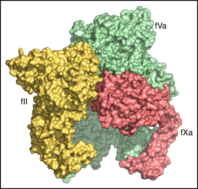
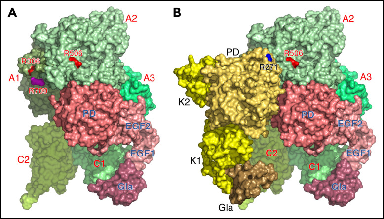
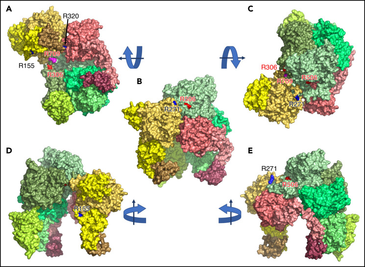
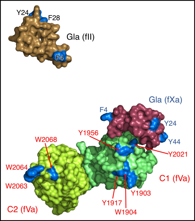
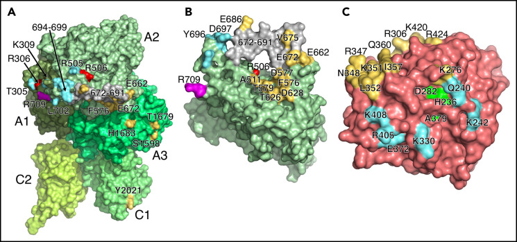
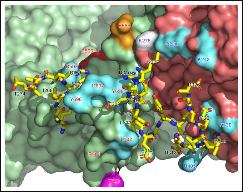
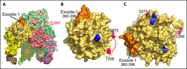
The intrinsic and extrinsic pathways of the coagulation cascade converge to a common step where the prothrombinase complex, comprising the enzyme factor Xa (fXa), the cofactor fVa, Ca2+ and phospholipids, activates the zymogen prothrombin to the protease thrombin. The reaction entails cleavage at 2 sites, R271 and R320, generating the intermediates prethrombin 2 and meizothrombin, respectively. The molecular basis of these interactions that are central to hemostasis remains elusive. We solved 2 cryogenic electron microscopy (cryo-EM) structures of the fVa-fXa complex, 1 free on nanodiscs at 5.3-Å resolution and the other bound to prothrombin at near atomic 4.1-Å resolution. In the prothrombin-fVa-fXa complex, the Gla domains of fXa and prothrombin align on a plane with the C1 and C2 domains of fVa for interaction with membranes. Prothrombin and fXa emerge from this plane in curved conformations that bring their protease domains in contact with each other against the A2 domain of fVa. The 672ESTVMATRKMHDRLEPEDEE691 segment of the A2 domain closes on the protease domain of fXa like a lid to fix orientation of the active site. The 696YDYQNRL702 segment binds to prothrombin and establishes the pathway of activation by sequestering R271 against D697 and directing R320 toward the active site of fXa. The cryo-EM structure provides a molecular view of prothrombin activation along the meizothrombin pathway and suggests a mechanism for cleavage at the alternative R271 site. The findings advance our basic knowledge of a key step of coagulation and bear broad relevance to other interactions in the blood.


Figures

None
Graphical abstract
Figure 1.
Figure 1.
Cryo-EM structures of prothrombinase free and bound to prothrombin. (A) The structure was solved at 5.3-Å resolution and shows the 2 proteins of the prothrombinase complex, fVa and fXa, in surface representation. Images including the nanodiscs are given in supplemental Figures 1 and 2. The architecture of the A1 (smudge), A2 (pale green), A3 (lime green), C1 (lime), and C2 (lemon) domains is solved in its entirety, except for the N-terminal 1546SNNGNRRNYY1555 sequence of the A3 domain immediately downstream of the site of thrombin activation at R1545. The overall arrangement of fVa is very similar to that of the free form solved recently. The architecture of fXa is fully resolved and provides a picture of the complete arrangement of the constitutive Gla (raspberry), EGF1 (dark salmon), EGF2 (salmon), and protease (deep salmon). The bound fXa features an overall conformation with the domains not vertically aligned but bent over the EGF1-EGF2 junction, as documented in several X-ray structures, and additionally 90° at the EGF1-Gla domain junction (supplemental Figure 4C). Also shown are the sites of fVa inactivation by activated protein C at R306 and R506 (red) and the site of thrombin activation at R709 (magenta). The enzyme is positioned in the complex along the A2, A3, and C1 domains of fVa, with the Gla domain aligned with C1 domain on the plane of the nanodisc (see supplemental Figure 1A). A segment of the A2 domain lowers on the fXa to fix the protease domain for optimal interaction with an incoming substrate (see Figure 2 for details). (B) The structure was solved at 4.1-Å resolution and shows fVa and fXa in the same arrangement found on nanodiscs (see panel A), which supports the ternary complex as a genuine representation of the prothrombin-prothrombinase complex. The constitutive domains of prothrombin are colored in sand (Gla domain), pale yellow (EGF1), yellow (EGF2), and orange yellow (protease domain, PD), with the Gla domain aligned with the homologous domain of fXa and the C1 and C2 domains of fVa for interaction with membranes. Prothrombin faces fXa in the complex and is positioned along the A2, A1, and C2 domains of fVa. Visible in this orientation is the site of activation at R271 (blue), located above the protease domain of fXa, but not R320 that inserts into the active site of fXa (see Figure 2 for details). The overall architecture of prothrombin is similar to that of the closed form (supplemental Figure 4A).
Figure 2.
Figure 2.
Cryo-EM structure of prothrombin bound to fVa and fXa. Molecular surface representation of the ternary complex of prothrombin fVa and fXa. The complex in Figure 1B is shown in different orientations obtained by 90° rotation of the reference view (B) as indicated by arrows. The constitutive domains of prothrombin, fVa and fXa are rendered in the same colors as in Figure 1B. Prothrombin engages prothrombinase through the protease domain (B,D-E) that binds to the A2 domain of fVa (B-D) and the protease domain of fXa (A-C). The protease domain of fXa is in close contact with the A2 domain of fVa (B,E). Labeled are the sites of prothrombin activation by prothrombinase at R271 (B-C,E) and R320, which is visible only looking up from the plane of the membrane (A) as it penetrates the active site of fXa. The preferred site of prothrombin activation in the absence of fVa, R155, is located on the side of prothrombin opposite to that facing fXa (D). The 672 to 691 region of the A2 domain of fVa changes conformation in the complex relative to the free form (C,E) and closes like a lid on the protease domain of fXa while making direct contacts with R271 of prothrombin (C,E; see also Figure 5 for details).
Figure 3.
Figure 3.
Membrane binding module of the prothrombin-prothrombinase complex. The Gla domains of prothrombin and fXa, along with the C1 and C2 domains of fVa, are colored as in Figure 1 and are shown in the same orientation as Figure 2A, that is, as seen from the plane of the membrane. The module features the Gla domain of fXa close, but not in contact, with the C1 domain of fVa, which is positioned relative to the C2 domain as seen in free fVa. The Gla domain of prothrombin, conversely, is widely separated from the Gla domain of fXa and the C2 domain of fVa. The module has an overall triangular arrangement with sides of up to 90 Å (Gla domain of prothrombin from Gla domain of fXa), 70 Å (Gla domain of fXa from C2 domain of fVa), and 110 Å (Gla domain of prothrombin from C2 domain of fVa). Residues likely involved in direct interaction with the membrane are highlighted (blue).
Figure 4.
Figure 4.
Interaction of fVa with fXa in prothrombinase. (A) Overall structure of fVa in surface representation, oriented as in Figures 1B and 2B, showing the residues involved in recognition of fXa (yellow) and prothrombin (cyan). The interaction with fXa involves mainly the A2 domain and few contacts in the A3 and C1 domains (Table 2). Residues in the 694 to 699 region (Table 2) are labeled as a group. The entire 672 to 691 segment (gray) moves >7 Å relative to the position in free fV to close like a lid over the protease domain of fXa. Also shown are the sites of inactivation by activated protein C at R306 and R506 (red) and the site of thrombin activation of fV at R709 (purple). (B) Surface representation of the A2 domain of fVa oriented as in Figure 2A, with all other domains, fXa and prothrombin removed for clarity. The view reveals the individual residues of fVa important for fXa binding (yellow) and their position relative to the 672 to 691 segment (gray) and the epitopes for prothrombin binding (cyan). (C) Surface representation of the protease domain of fXa oriented as in Figure 2D and rotated 30° along the x-axis, with all other domains, fVa and prothrombin removed for clarity. Residues of the catalytic triad (green), with S379 replaced by Ala, are in the center. The view reveals the residues involved in fVa binding (yellow) as being located toward the C-terminal helix (K420, R434) and the 340 to 350 (c165-175) segment of the protease domain. Also shown are residues responsible for binding of prothrombin (cyan) around the active site entrance (Q240, K242, K33) and the Na+ site region (E372, R405, K408). Particularly important is the strong electrostatic coupling of R347 of fXa with E572 and E662 of fVa, as identified by biochemical studies.
Figure 5.
Figure 5.
Interaction of prothrombin with prothrombinase. Molecular surface of interaction between the A2 domain of fVa (pale green surface), fXa (deep salmon surface), and prothrombin (yellow sticks) that details how fVa orchestrates preferential binding of R320 to the active site of fXa. Shown is the segment 269EGRTAT274 comprising the site of cleavage at R271 and the longer segment 307KTERELLESYIDGRIVEGSD326 comprising the site of cleavage at R320. Relevant epitopes of fVa and fXa for interaction with prothrombin are colored in cyan. The interaction between E686 of fVa (orange) with K276 of fXa (white) is also labeled, along with R506 and R709 of fVa. The segment 696YDYQNRL702 separates the 2 prothrombin segments and sequesters R271 with a strong ionic interaction with D697 fixed by a nearby interaction between E269 and R505 of fVa. The segment then organizes the proximal portion of the R320 site for interaction with the active site of fXa. Flanking D697 are Y696 in hydrophobic interaction with F535 and Y698 coupled to R310 through a cation-π interaction and in hydrophobic contact with L312. The fragment then turns toward fXa where strong electrostatic interactions involve D318 at P3, E323 at P3′ and D326 at P6’ with residues R405, K370, and K242 of fXa, respectively. An additional ionic interaction is established between K307 of prothrombin and Q240 of fXa. As a result of these interactions, R320 penetrates the active site of fXa to initiate activation along the meizothrombin pathway.
Figure 6.
Figure 6.
Putative interaction of prothrombin in the open form with prothrombinase. (A) The complex was obtained by overlaying the structure of prothrombin in the open form (PDB ID 5EDM) to the closed form in the cryo-EM structure (Figures 1B and 2). The open form aligns the Gla domain with the homologous domain of fXa and the C1 and C2 domains of fVa. Unfortunately, the entire segment 257GDGLDEDSDRAIEGRTAT274 containing the site of cleavage at R271 was not resolved in the 5EDM structure (B, dotted lines). Residue R320 moves 18 Å upward and clashes with the area of contact between E686 of fVa and K376 of fXa (yellow circle; see also Figure 5). A movement of the entire 672 to 691 region (gray) of the A2 domain would be necessary to accommodate the protease domain of prothrombin in the open form. Exosite-1 of prothrombin (residues 382RIGKHSRTRYERNIE396, orange) moves closer to but not in contact with fVa. (B-C) Protease domain of prothrombin in the open (B) and closed (C) forms obtained after rotation of panel A 90° clockwise along the y-axis (B) or directly from Figure 2E (C), with fVa, fXa, and the auxiliary domains (EGF1, EGF2, Gla) removed for clarity. Transition from the closed to the open form moves exosite 1 closer to fVa and relocates R320 18 Å upward from the position in the closed form (indicated in panel B by a yellow circle, for reference). The transition also causes a significant clockwise rotation of the entire segment 257 to 274 containing the R271 site (C), not visible in the open form (B, dotted lines), with the Cα-Cα distance between T256 and S275 shrinking from 37 to 16 Å. The rotation would bring R271 closer to the active site of fXa in the open form.

Comment in

  • Shining a light on thrombin activation.
    Spiegel PC. Spiegel PC. Blood. 2022 Jun 16;139(24):3451-3453. doi: 10.1182/blood.2022016537. Blood. 2022. PMID: 35708725 No abstract available.
  • What the neighbors say.
    [No authors listed] [No authors listed] J Thromb Haemost. 2022 Aug;20(8):1755. doi: 10.1111/jth.15796. J Thromb Haemost. 2022. PMID: 35859280 No abstract available.

References

    1. Davie EW, Fujikawa K, Kisiel W. The coagulation cascade: Initiation, maintenance, and regulation. Biochemistry. 1991;30(43):10363-10370. - PubMed
    1. Mann KG. Thrombin formation. Chest. 2003;124(3 suppl):4S-10S. - PubMed
    1. Gailani D, Broze GJ Jr. Factor XI activation in a revised model of blood coagulation. Science. 1991;253(5022):909-912. - PubMed
    1. Rosing J, Tans G, Govers-Riemslag JW, Zwaal RF, Hemker HC. The role of phospholipids and factor Va in the prothrombinase complex. J Biol Chem. 1980;255(1):274-283. - PubMed
    1. Mann KG, Kalafatis M. Factor V: A combination of Dr Jekyll and Mr Hyde. Blood. 2003;101(1):20-30. - PubMed

Publication types