. 2022 Jul 15:255:119200.
doi: 10.1016/j.neuroimage.2022.119200. Epub 2022 Apr 13.

Ultra-high field (10.5T) diffusion-weighted MRI of the macaque brain

Affiliations

Ultra-high field (10.5T) diffusion-weighted MRI of the macaque brain

Mark D Grier et al. Neuroimage. .

Abstract

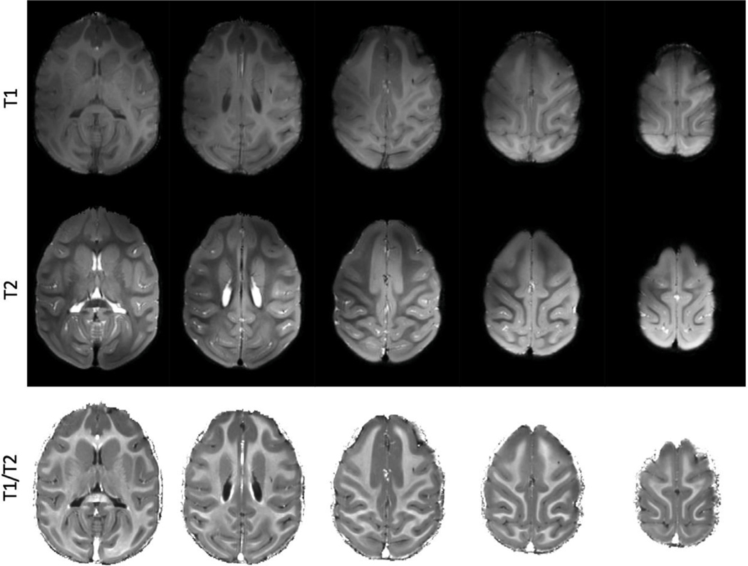
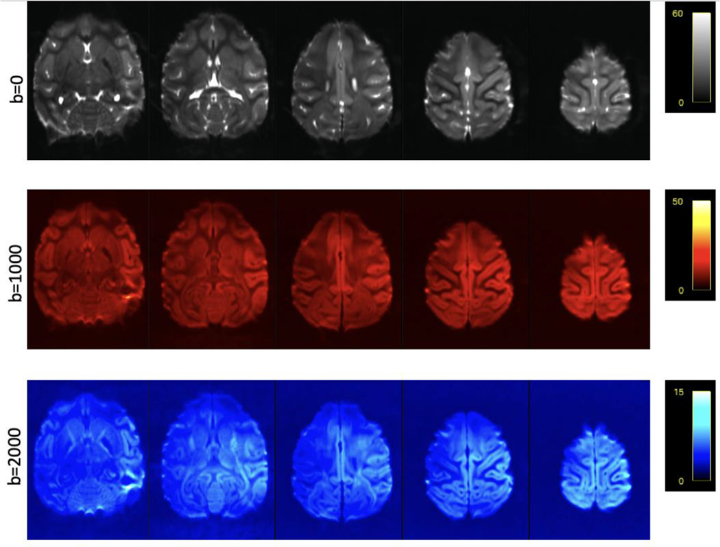
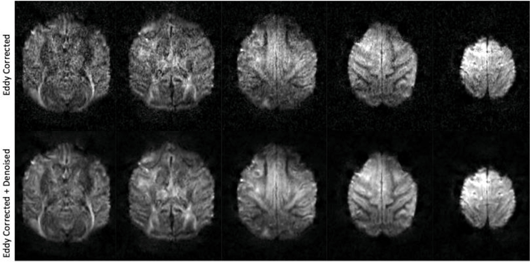
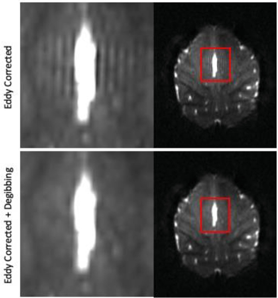
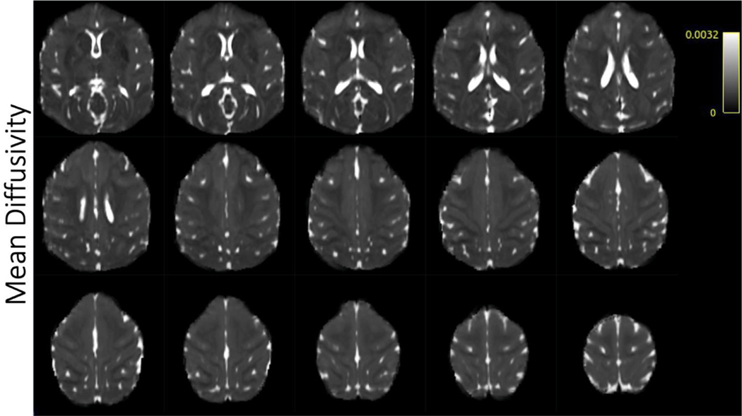
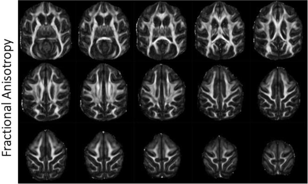
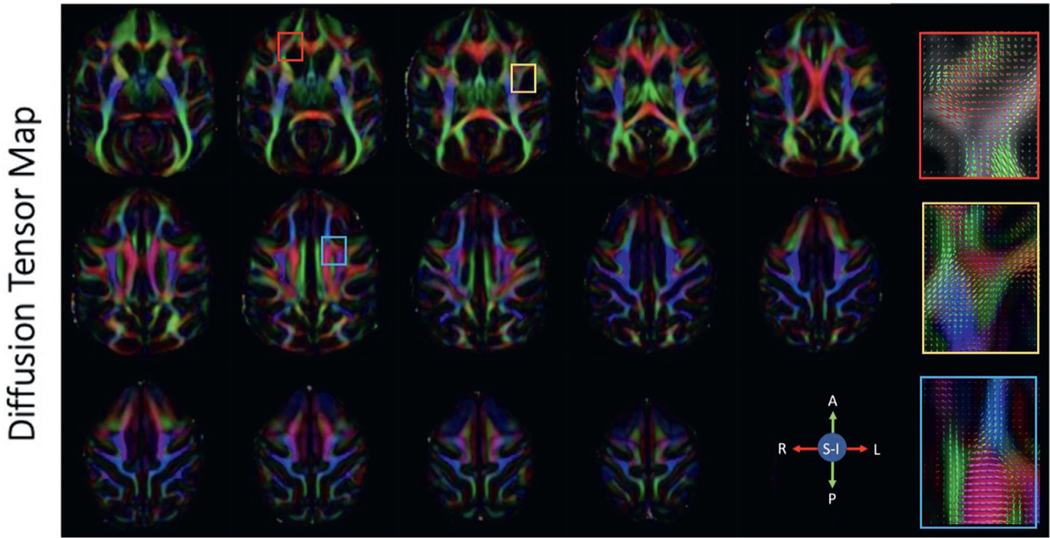
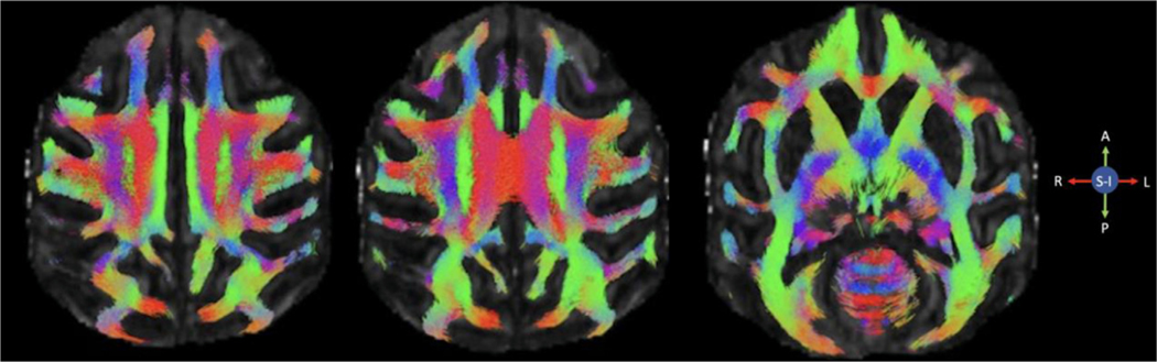
Diffu0sion-weighted magnetic resonance imaging (dMRI) is a non-invasive imaging technique that provides information about the barriers to the diffusion of water molecules in tissue. In the brain, this information can be used in several important ways, including to examine tissue abnormalities associated with brain disorders and to infer anatomical connectivity and the organization of white matter bundles through the use of tractography algorithms. However, dMRI also presents certain challenges. For example, historically, the biological validation of tractography models has shown only moderate correlations with anatomical connectivity as determined through invasive tract-tracing studies. Some of the factors contributing to such issues are low spatial resolution, low signal-to-noise ratios, and long scan times required for high-quality data, along with modeling challenges like complex fiber crossing patterns. Leveraging the capabilities provided by an ultra-high field scanner combined with denoising, we have acquired whole-brain, 0.58 mm isotropic resolution dMRI with a 2D-single shot echo planar imaging sequence on a 10.5 Tesla scanner in anesthetized macaques. These data produced high-quality tractograms and maps of scalar diffusion metrics in white matter. This work demonstrates the feasibility and motivation for in-vivo dMRI studies seeking to benefit from ultra-high fields.

Keywords: Diffusion MRI; Nonhuman primate; Structural connectivity; Tractography.


Figures

Fig. 1.
Fig. 1.. Structural images of the macaque brain acquired at 10.5T.
Top panel shows T1w images. Middle panel shows T2w images. The bottom panel shows a T1w/T2w ratio image.
Fig. 2.
Fig. 2.. Signal-to-noise ratio maps for 0.58 mm isotropic resolution dMRI data acquired at 10.5T from a single subject, I.
The average SNR for each shell is shown. Selected axial slices show a relatively homogenous SNR through the entire brain.
Fig. 3.
Fig. 3.. Effect of MPPCA denoising on diffusion weighted images.
The top panel shows 5 axial slices of data processed through Eddy and Topup. The bottom panel shows Eddy corrected with additional denoising.
Fig. 4.
Fig. 4.. Removal of Gibbs ringing artifact.
The top panel shows parallel lines in the b = 0 image immediately adjacent to a ventricle/gray matter interface. The bottom panel shows the data after being processed with mrdegibbs. The ringing artifact is removed, while retaining the integrity of the original data.
Fig. 5.
Fig. 5.
Selected axial slices showing mean diffusivity in a single subject acquired at 0.58 mm isotropic resolution.
Fig. 6.
Fig. 6.. Fractional anisotropy.
Selected axial slices showing fractional anisotropy in a single subject acquired at 0.58 mm isotropic resolution.
Fig. 7.
Fig. 7.. Diffusion tensor maps.
Selected axial slices showing the principal diffusion direction in a single subject acquired at 0.58 mm isotropic resolution. Red, green and blue represent fibers traveling right-left, anterior-posterior and superior-inferior respectively. The colored boxes correspond to the fiber orientation distribution functions displayed on the right.
Fig. 8.
Fig. 8.. Representative whole brain tractograms from a single subject.
Selected axial slices showing whole brain streamlines.
Fig. 9.
Fig. 9.. Tractography in dMRI data collected at 10.5T.
Tractography from a single subject with seeds in the motor cortex (A), caudal corpus callosum (B), precuneus (C), and ventrolateral prefrontal cortex (D). Streamlines are overlaid on a T1 image registered to the diffusion space.
Fig. 10.
Fig. 10.
U-fibers. Fractional anisotropy maps for the arcuate and intraparietal sulci that contain short association fibers, also known as U-fibers. The middle panel shows the ODFs in the corresponding region. The lower panel displays streamlines generated from a seed that straddles the gray matter/white matter boundary as described by Reveley (Reveley et al., 2015a).

References

    1. Alexander AL, Hasan KM, Lazar M, Tsuruda JS, Parker DL, 2001. Analysis of partial volume effects in diffusion-tensor MRI. Magn. Reson. Med doi: 10.1002/mrm.1105. - DOI - PubMed
    1. Alomair OI, Brereton IM, Smith MT, Galloway GJ, Kurniawan ND, 2015. In vivo high angular resolution diffusion-weighted imaging of mouse brain at 16.4 Tesla. PLOS ONE. doi: 10.1371/journal.pone.0130133. - DOI - PMC - PubMed
    1. Andersson JLR, Graham MS, Zsoldos E, Sotiropoulos SN, 2016. Incorporating outlier detection and replacement into a non-parametric framework for movement and distortion correction of diffusion MR images. Neuroimage 141, 556–572. - PubMed
    1. Andersson JLR, Skare S, Ashburner J, 2003. How to correct susceptibility distortions in spin-echo echo-planar images: application to diffusion tensor imaging. Neuroimage 20, 870–888. - PubMed
    1. Andersson JLR, Sotiropoulos SN, 2016. An integrated approach to correction for off-resonance effects and subject movement in diffusion MR imaging. Neuroimage 125, 1063–1078. - PMC - PubMed

Publication types