

Corticomedullary shunting after ischaemia and reperfusion in the porcine kidney?
- PMID: 35428270
- PMCID: PMC9013123
- DOI: 10.1186/s12882-022-02780-0
Corticomedullary shunting after ischaemia and reperfusion in the porcine kidney?
Abstract
Background: Renal perfusion may redistribute from cortex to medulla during systemic hypovolaemia and after renal ischaemia for other reasons, but there is no consensus on this matter. We studied renal perfusion after renal ischaemia and reperfusion.
Methods: Renal perfusion distribution was examined by use of 153Gadolinium-labeled microspheres (MS) after 2 h (hrs) and 4 h ischaemia of the pig kidney followed by 4 h of reperfusion. Intra-arterial injected MS are trapped in the glomeruli in renal cortex, which means that MS are not present in the medulla under normal physiological conditions.
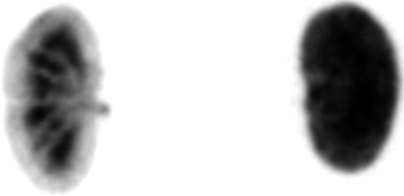
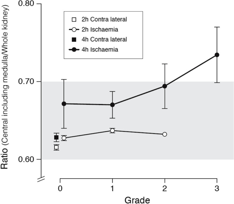
Results: Visual evaluation after reperfusion demonstrated that MS redistributed from the renal cortex to the medulla in 6 out of 16 pigs (38%) subjected to 4 h ischaemia and in one out of 18 pigs subjected to 2 h ischaemia. Central renal uptake of MS covering the medullary/total renal uptake was significantly higher in kidneys subjected to 4 h ischaemia compared with pigs subjected to 2 h ischaemia (69 ± 5% vs. 63 ± 1%, p < 0.001), and also significantly higher than in the contralateral kidney (69 ± 5% vs. 63 ± 2%, p < 0.001). Analysis of blood and urine demonstrated no presence of radioactivity.
Conclusion: The study demonstrated the presence of MS in the renal medulla in response to renal ischaemia and reperfusion suggesting that severe ischaemia and reperfusion of the pig kidney leads to opening of functional shunts bypassing glomeruli.
Keywords: Microspheres; Renal blood flow; Renal ischaemia; Renal perfusion; Renal redistribution; Renal shunts.
© 2022. The Author(s).
Conflict of interest statement
The authors declare that they have no competing interests.
Figures
References
-
- Munger K, Kost C, Jr, Brenner B, Maddox D. The renal circulations and glomerular ultrafiltration. In: Taal M, Chertow G, Marsden P, Skorecki K, Yu Y, Brenner B, editors. Brenner and Rectors, The Kidney. Philadelphia: Elsevier Saunders; 2012. pp. 94–137.
-
- Trueta J, Barclay A, Daniel P. Studies of the renal circulation. Oxford: Blackwell Scientific Publications Ltd; 1947.
-
- Daniel P, Peabody C, Prichard M. Cortical ischaemia of the kidney with maintained blood flow through the medulla. Q J Exp Physiol Cogn Med Sci. 1952;37:11–18. - PubMed
-
- Daniel P, Peabody C, Prichard M. Observations on the circulation through the cortex and the medulla of the kidney. Q J Exp Physiol Cogn Med Sc. 1951;36:199–203. - PubMed
Publication types
MeSH terms
LinkOut - more resources
Full Text Sources
