Multicenter Study
. 2022 Apr 15;26(1):110.
doi: 10.1186/s13054-022-03978-2.

Transcranial Doppler as a screening test to exclude intracranial hypertension in brain-injured patients: the IMPRESSIT-2 prospective multicenter international study

Affiliations
Multicenter Study

Transcranial Doppler as a screening test to exclude intracranial hypertension in brain-injured patients: the IMPRESSIT-2 prospective multicenter international study

Frank A Rasulo et al. Crit Care. .

Abstract

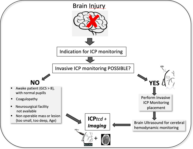
Background: Alternative noninvasive methods capable of excluding intracranial hypertension through use of transcranial Doppler (ICPtcd) in situations where invasive methods cannot be used or are not available would be useful during the management of acutely brain-injured patients. The objective of this study was to determine whether ICPtcd can be considered a reliable screening test compared to the reference standard method, invasive ICP monitoring (ICPi), in excluding the presence of intracranial hypertension.

Methods: This was a prospective, international, multicenter, unblinded, diagnostic accuracy study comparing the index test (ICPtcd) with a reference standard (ICPi), defined as the best available method for establishing the presence or absence of the condition of interest (i.e., intracranial hypertension). Acute brain-injured patients pertaining to one of four categories: traumatic brain injury (TBI), subarachnoid hemorrhage (SAH), intracerebral hemorrhage (ICH) or ischemic stroke (IS) requiring ICPi monitoring, were enrolled in 16 international intensive care units. ICPi measurements (reference test) were compared to simultaneous ICPtcd measurements (index test) at three different timepoints: before, immediately after and 2 to 3 h following ICPi catheter insertion. Sensitivity, specificity, positive (PPV) and negative predictive values (NPV) were calculated at three different ICPi thresholds (> 20, > 22 and > 25 mmHg) to assess ICPtcd as a bedside real-practice screening method. A receiver operating characteristic (ROC) curve analysis with the area under the curve (AUC) was used to evaluate the discriminative accuracy and predictive capability of ICPtcd.

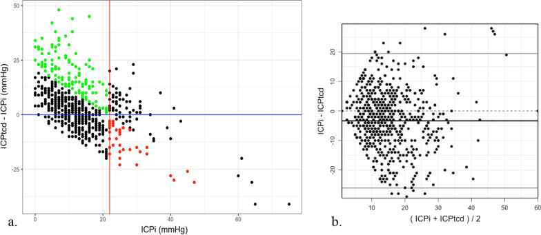
Results: Two hundred and sixty-two patients were recruited for final analysis. Intracranial hypertension (> 22 mmHg) occurred in 87 patients (33.2%). The total number of paired comparisons between ICPtcd and ICPi was 687. The NPV was elevated (ICP > 20 mmHg = 91.3%, > 22 mmHg = 95.6%, > 25 mmHg = 98.6%), indicating high discriminant accuracy of ICPtcd in excluding intracranial hypertension. Concordance correlation between ICPtcd and ICPi was 33.3% (95% CI 25.6-40.5%), and Bland-Altman showed a mean bias of -3.3 mmHg. The optimal ICPtcd threshold for ruling out intracranial hypertension was 20.5 mmHg, corresponding to a sensitivity of 70% (95% CI 40.7-92.6%) and a specificity of 72% (95% CI 51.9-94.0%) with an AUC of 76% (95% CI 65.6-85.5%).

Conclusions and relevance: ICPtcd has a high NPV in ruling out intracranial hypertension and may be useful to clinicians in situations where invasive methods cannot be used or not available.

Trial registration: NCT02322970 .

Keywords: Brain injury; Intracranial hypertension; Intracranial pressure; Noninvasive monitoring.


Conflict of interest statement

The authors declare that they have no competing interests.

Figures

Fig. 1
Fig. 1
The three time frames (T1, T2, T3) of paired invasive (ICPi) and noninvasive (ICPtcd) measurements of intracranial pressure (ICP). At TIME 1 (T1), ICPtcd was obtained shortly before performing the burr hole procedure and was compared to the first ICPi reading once the invasive probe was positioned. At TIME 2 (T2) and TIME 3 (T3), paired ICPtcd and ICPi were obtained immediately after TIME 1 and 2 to 3 h following the second reading
Fig. 2
Fig. 2
Patient recruitment plus the Standards for Reporting of Diagnostic Accuracy (STARD) flowchart
Fig. 3
Fig. 3
Percentages of patients with intracranial hypertension (IH) and negative (NPV) and positive (PPV) predictive values at the three intracranial pressure (ICP) thresholds. a Percentage of patients with ICP above (red silhouette indicating IH) and below (green silhouette indicating normal ICP) the given ICP thresholds of 20, 22 and 25 mmHg. b NPV and PPV (%) of the averaged time frames (T1,2,3) at the three ICP thresholds (20, 22 and 25 mmHg). NPV = green silhouette indicating true negatives and grey silhouette indicating false negatives. PPV = red silhouette indicating true positives and grey silhouette indicating false positives
Fig. 4
Fig. 4
a Distribution of differences between paired ICPtcd and ICPi measurements as a function of ICPi. Black points represent concordant measurements (either true positive and true negative cases); green points indicate ICPi < 22 and ICPtcd ≥ 22 (false positive measurements); red points indicate ICPi ≥ 22 and ICPtcd < 22 (false negative cases). b Bland–Altman analysis yielded a mean bias (ICPtcd–ICPi) of − 3.3 mmHg with an agreement range comprised between − 26.1 and + 19.5
Fig. 5
Fig. 5
Algorithm for noninvasive intracranial pressure monitoring through the use of transcranial Doppler (ICPtcd). Once indication for ICP monitoring is decided, application of the invasive method (gold standard) is evaluated: If NO (invasive ICP monitoring not possible) for the presence of one or more of the reasons provided, ICPtcd associated with brain imaging should be performed. *The result should not act as a deterrent for transferal towards a hospital with a Neurosurgical facility or to perform further brain imaging studies, instead ICPtcd should be used as an adjunct capable of providing valuable information for the Clinician; if YES (invasive ICP monitoring possible), then burr hole and insertion of a catheter within the brain parenchyma should be performed. *However, brain ultrasound with transcranial Doppler may be useful in order to obtain surrogate information regarding brain hemodynamics through the evaluation of cerebral blood flow velocity

References

    1. Carney N, Totten AM, O’Reilly C, et al. Guidelines for the management of severe traumatic brain injury, fourth edition. Neurosurgery. 2017;80(1):6–15. doi: 10.1227/NEU.0000000000001432. - DOI - PubMed
    1. Güiza F, Depreitere B, Piper I, et al. Visualizing the pressure and time burden of intracranial hypertension in adult and paediatric traumatic brain injury. Intensive Care Med. 2015;41(6):1067–1076. doi: 10.1007/s00134-015-3806-1. - DOI - PubMed
    1. Livesay S, Fried H, Gagnon D, et al. clinical performance measures for neurocritical care: a statement for healthcare professionals from the Neurocritical Care Society. Neurocrit Care. 2020;32:5–79. doi: 10.1007/s12028-019-00846-w. - DOI - PubMed
    1. Barber MA, Helmer SD, Morgan JT, Haan JM. Placement of intracranial pressure monitors by non-neurosurgeons: excellent outcomes can be achieved. J Trauma Acute Care Surg. 2012;73(3):558–565. doi: 10.1097/TA.0b013e318265cb75. - DOI - PubMed
    1. Jinadasa SP, Ruan QZ, Bayoumi AB, et al. Hemorrhagic complications of invasive intracranial pressure monitor placement in acute liver failure: outcomes of a single-center protocol and comprehensive literature review. Neurocrit Care. 2021;35(1):87–102. doi: 10.1007/s12028-020-01143-7. - DOI - PubMed

Publication types

Associated data