

Optical coherence tomography as retinal imaging biomarker of neuroinflammation/neurodegeneration in systemic disorders in adults and children
- PMID: 35428871
- PMCID: PMC9012155
- DOI: 10.1038/s41433-022-02056-9
Optical coherence tomography as retinal imaging biomarker of neuroinflammation/neurodegeneration in systemic disorders in adults and children
Erratum in
-
Correction: Optical coherence tomography as retinal imaging biomarker of neuroinflammation/neurodegeneration in systemic disorders in adults and children.Eye (Lond). 2023 Feb;37(2):379. doi: 10.1038/s41433-022-02083-6. Eye (Lond). 2023. PMID: 35550606 Free PMC article. No abstract available.
Abstract
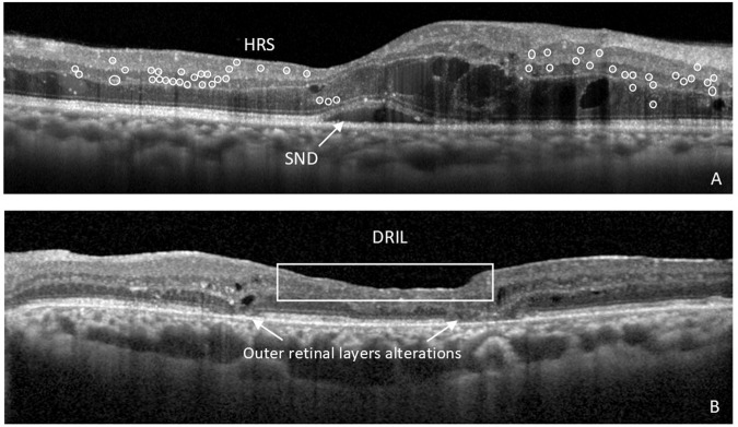
The retina and the optic nerve are considered extensions of the central nervous system (CNS) and thus can serve as the window for evaluation of CNS disorders. Spectral domain optical coherence tomography (OCT) allows for detailed evaluation of the retina and the optic nerve. OCT can non-invasively document changes in single retina layer thickness and structure due to neuronal and retinal glial cells (RGC) modifications in systemic and local inflammatory and neurodegenerative diseases. These can include evaluation of retinal nerve fibre layer and ganglion cell complex, hyper-reflective retinal spots (HRS, sign of activated microglial cells in the retina), subfoveal neuroretinal detachment, disorganization of the inner retinal layers (DRIL), thickness and integrity of the outer retinal layers and choroidal thickness. This review paper will report the most recent data on the use of OCT as a non invasive imaging biomarker for evaluation of the most common systemic neuroinflammatory and neurodegenerative/neurocognitive disorders in the adults and in paediatric population. In the adult population the main focus will be on diabetes mellitus, multiple sclerosis, optic neuromyelitis, neuromyelitis optica spectrum disorders, longitudinal extensive transverse myelitis, Alzheimer and Parkinson diseases, Amyotrophic lateral sclerosis, Huntington's disease and schizophrenia. In the paediatric population, demyelinating diseases, lysosomal storage diseases, Nieman Pick type C disease, hypoxic ischaemic encephalopathy, human immunodeficiency virus, leukodystrophies spinocerebellar ataxia will be addressed.
摘要: 视网膜和视神经是中枢神经系统 (CNS) 的延续, 因此可以作为评估CNS疾病的窗口。频域光学相干断层扫描 (SD-OCT) 可以对视网膜和视神经进行详细的评估。OCT可以无创性地记录系统性和局部炎症/神经退行性病变中, 由于神经元和视网膜胶质细胞 (RGC) 改变引起的视网膜单层厚度和结构的变化。OCT的观察的指征包括评估视网膜神经纤维层和神经节细胞复合体、视网膜高反射点 (HRS, 视网膜中小胶质细胞激活的征象) 、中心凹下神经视网膜脱离、视网膜内层结构紊乱 (DRIL) 、视网膜外层的厚度和完整性以及脉络膜厚度。本文将总结OCT作为无创成像生物标志物评估成人和儿童中最常见的系统性神经炎症和神经退行性病变/神经认知障碍的最新数据。在成人中, 我们最关注的疾病为糖尿病、多发性硬化症、视神经脊髓炎、视神经脊髓炎谱系障碍、纵向广泛横贯性脊髓炎、阿尔茨海默病和帕金森病、肌萎缩侧索硬化症、亨廷顿病和精神分裂症。在儿童中, 我们着重讨论的疾病有脱髓鞘疾病、溶酶体贮积病、尼曼-匹克病、缺氧缺血性脑病、人类免疫缺陷病毒、脑白质营养不良脊髓小脑性共济失调。.
© 2022. The Author(s), under exclusive licence to The Royal College of Ophthalmologists.
Conflict of interest statement
SV: Abbvie-Allergan, Apellis, Bayer, Novartis, Roche SPA: Advisory Board participation MMP: none MEH: This work was supported by the National Institutes of Health EY014800 and an Unrestricted Grant from Research to Prevent Blindness, Inc., New York, NY, to the Department of Ophthalmology & Visual Sciences, University of Utah; and the National Institutes of Health R01EY015130 and R01EY017011 to MEH. LO’T: Bayer and Novartis: Advisory Board participation. AN: none. Celeste Limoli: none. EV: Allergan: Consultant/Advisor, Lecture fees, Grant support (AAO Financial Disclosure and First-slide Policy 2021); FB Vision and Santen: Consultant/Advisor, Lecture fees; Bruschettini, Oftagest, Sooft, Thea and Visufarma: Lecture fees; Alfa intes, OffHealth, Servimed and Shire: Grant support (AAO Financial Disclosure and First-slide Policy 2021). PN: none.
Figures
Comment in
-
Hyperreflective spots in cerebral malaria.Eye (Lond). 2023 Oct;37(15):3295. doi: 10.1038/s41433-023-02485-0. Epub 2023 Mar 10. Eye (Lond). 2023. PMID: 36899110 Free PMC article. No abstract available.
References
-
- Bakhtadze S, Geladze N, Khachapuridze N. Inflammation in childhood epilepsy syndromes. Georgian Med N. 2021;312:88–92. - PubMed
Publication types
MeSH terms
Substances
Grants and funding
LinkOut - more resources
Full Text Sources
