Review
. 2022 Apr 16;79(5):243.
doi: 10.1007/s00018-022-04278-2.

The role of bile acids in carcinogenesis

Affiliations
Review

The role of bile acids in carcinogenesis

Tadeja Režen et al. Cell Mol Life Sci. .

Abstract

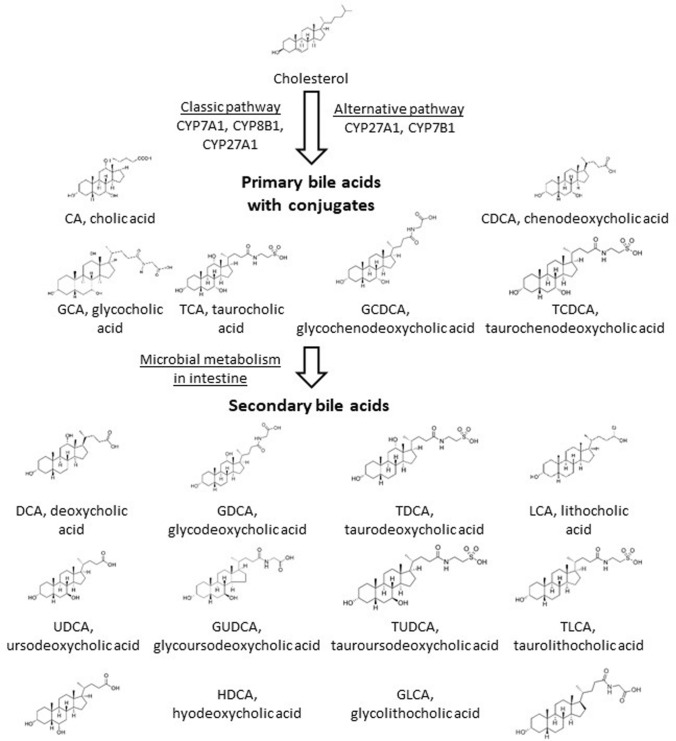
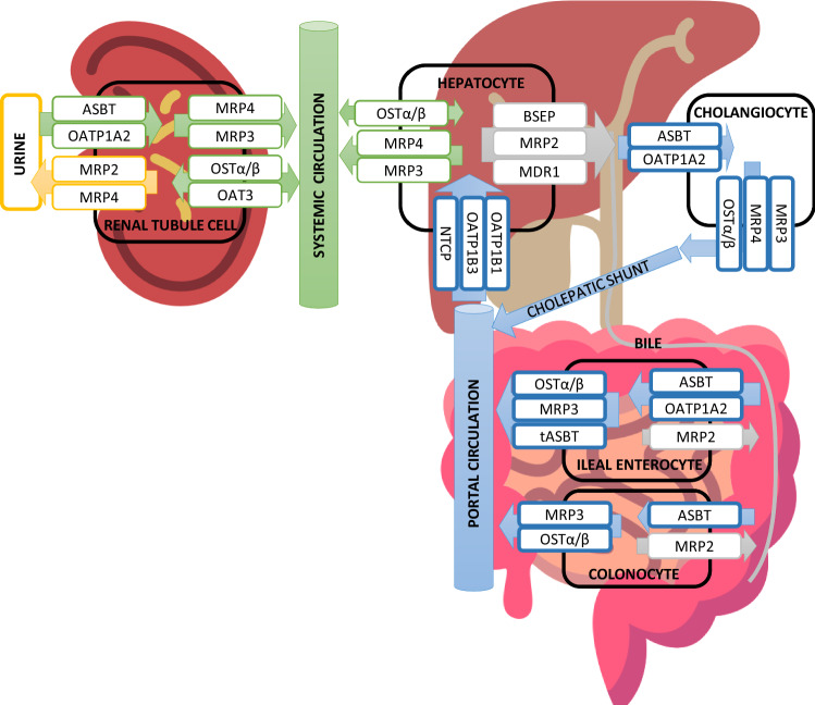
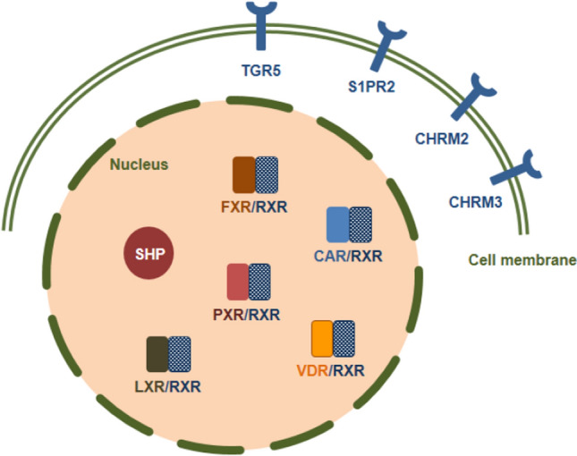
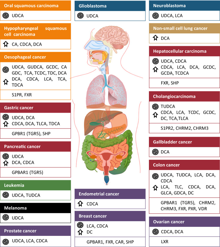
Bile acids are soluble derivatives of cholesterol produced in the liver that subsequently undergo bacterial transformation yielding a diverse array of metabolites. The bulk of bile acid synthesis takes place in the liver yielding primary bile acids; however, other tissues have also the capacity to generate bile acids (e.g. ovaries). Hepatic bile acids are then transported to bile and are subsequently released into the intestines. In the large intestine, a fraction of primary bile acids is converted to secondary bile acids by gut bacteria. The majority of the intestinal bile acids undergo reuptake and return to the liver. A small fraction of secondary and primary bile acids remains in the circulation and exert receptor-mediated and pure chemical effects (e.g. acidic bile in oesophageal cancer) on cancer cells. In this review, we assess how changes to bile acid biosynthesis, bile acid flux and local bile acid concentration modulate the behavior of different cancers. Here, we present in-depth the involvement of bile acids in oesophageal, gastric, hepatocellular, pancreatic, colorectal, breast, prostate, ovarian cancer. Previous studies often used bile acids in supraphysiological concentration, sometimes in concentrations 1000 times higher than the highest reported tissue or serum concentrations likely eliciting unspecific effects, a practice that we advocate against in this review. Furthermore, we show that, although bile acids were classically considered as pro-carcinogenic agents (e.g. oesophageal cancer), the dogma that switch, as lower concentrations of bile acids that correspond to their serum or tissue reference concentration possess anticancer activity in a subset of cancers. Differences in the response of cancers to bile acids lie in the differential expression of bile acid receptors between cancers (e.g. FXR vs. TGR5). UDCA, a bile acid that is sold as a generic medication against cholestasis or biliary surge, and its conjugates were identified with almost purely anticancer features suggesting a possibility for drug repurposing. Taken together, bile acids were considered as tumor inducers or tumor promoter molecules; nevertheless, in certain cancers, like breast cancer, bile acids in their reference concentrations may act as tumor suppressors suggesting a Janus-faced nature of bile acids in carcinogenesis.

Keywords: Bile acid; Bile acid biosynthesis; Bile acid receptors; Bile acid transporters; Breast cancer; CA; CAR; CDCA; Carcinogenesis; Colorectal carcinoma; DCA; Epithelial–mesenchymal transition; FXR; Gastric cancer; Hepatocellular carcinoma; LCA; LXR; Microbiome; Muscarinic receptor CHRM2; Muscarinic receptor CHRM3; Oesophageal carcinoma; Ovarian cancer; Oxidative stress; PXR; Pancreatic adenocarcinoma; Primary bile acid; Prostate cancer; S1PR2; SHP; Secondary bile acid; TGR5; UDCA; VDR; Warburg metabolism.


Conflict of interest statement

The authors declare no conflict of interest.

Figures

Fig. 1
Fig. 1
Scheme of the classical and alternative bile acids in humans. Only enzymes of the CYP family are listed while the pathway involves enzymes of other protein families. CA and DCA are conjugated and further metabolized in the intestine
Fig. 2
Fig. 2
A scheme of enterohepatic and systemic circulation of bile acids and the transporters in different human cells. Transporters are coloured according to which part of the circulation they belong to. Blue are efflux and influx transporters, which transport BAs in portal circulation. Grey are efflux transporters, which contribute to bile export into bile and faeces. Green are transporters, which are responsible for BA transport into the systemic circulation. Yellow are transporters involved in the efflux of BAs into urine. ASBT/SLC10A2 sodium-dependent bile acid transporter, BSEP/ABCB11 ATP-dependent cassette transporter, MRP2/ABCC2 multidrug resistance-associated protein 2, MRP3/ABCC3 multidrug resistance-associated protein 3, MRP4/ABCC4 multidrug resistance-associated protein 4, OATP1A2/SLCO1A2 Solute Carrier Organic Anion Transporter Family Member 1A2, OATP1B/SLCO1B Solute Carrier Organic Anion Transporter Family, SLC51A/B or OSTα/β Solute Carrier Family members, SLC10A2/ASBT sodium-dependent bile acid transporter
Fig. 3
Fig. 3
The subcellular localization of bile acid receptors. TGR5 G protein-coupled bile acid receptor 1, S1PR2 Sphingosine-1-phosphate receptor 2, CHRM2 Muscarinic receptor-2, CHRM3 Muscarinic receptor-3, FXR Farnesoid X receptor, PXR Pregnane X receptor, CAR Constitutive androstane receptor, VDR Vitamin D receptor, SHP Small heterodimer partner
Fig. 4
Fig. 4
Different roles of bile acids and bile acids receptors in a wide variety of cancers. Some BAs have opposite effects, which depend on the cell line, BA concentration and other treatment conditions. The crossed circle symbol marks the tumor suppressor effects and the arrow marks the tumor promoter effects. CA Cholic acid, CAR Constititive androstane receptor, CDCA Chenodeoxycholic acid, CHRM2/M3 Muscarinic receptor 2 and 3, DC Deoxycholate, DCA Deoxycholic acid, FXR Farnesoid X receptor, GCDA Glycochenodeoxycholate acid, GCDC Glycochenodeoxycholate, GDC Glycodeoxycholate, GDCA Glycodeoxycholic acid, GLCA Glycolithocholic acid, GUDCA Glycoursodeoxycholic acid, LCA Lithocholic acid, PXR Pregnane X receptor, S1PR2 Sphingosine-1-phosphate receptor 2, SHP Small heterodimer partner, TCA Taurocholic acid, TCDC Taurochenodeoxycholate, TCDCA Taurochenodeoxycholic acid, TDC Taurodeoxycholate, TDCA Taurodeoxycholic acid, TGR5/GPBAR1 G protein- coupled bile acid receptor 1, TLC Taurolithocholate, TLCA Taurolithocholic acid, TUDCA Tauroursodeoxycholic acid, UDCA Ursodeoxycholic acid, VDR Vitamin D receptor

References

    1. Stieger B. Biliary cholesterol secretion: more lessons from plants? J Hepatol. 2003;38:843–846. doi: 10.1016/S0168-8278(03)00194-6. - DOI - PubMed
    1. Pellicciari R, Gioiello A, Costantino G. Potential therapeutic applications of farnesoid X receptor (FXR) modulators. Expert Opin Ther Pat. 2006;16:333–341. doi: 10.1517/13543776.16.3.333. - DOI
    1. Cai X, Young GM, Xie W. The xenobiotic receptors PXR and CAR in liver physiology, an update. Biochim Biophys Acta Mol Basis Dis. 2021;1867:166101. doi: 10.1016/j.bbadis.2021.166101. - DOI - PMC - PubMed
    1. Sipos A, Ujlaki G, Mikó E, Maka E, Szabó J, Uray K, Krasznai Z, Bai P. The role of the microbiome in ovarian cancer: mechanistic insights into oncobiosis and to bacterial metabolite signaling. Mol Med. 2021;27:33. doi: 10.1186/s10020-021-00295-2. - DOI - PMC - PubMed
    1. Kiss B, Mikó E, Sebő É, Toth J, Ujlaki G, Szabó J, Uray K, Bai P, Árkosy P. Oncobiosis and microbial metabolite signaling in pancreatic adenocarcinoma. Cancers (Basel) 2020;12:E1068. doi: 10.3390/cancers12051068. - DOI - PMC - PubMed

Substances