New clues to understand gastroschisis. Embryology, pathogenesis and epidemiology
- PMID: 35431359
- PMCID: PMC8973314
- DOI: 10.25100/cm.v52i3.4227
New clues to understand gastroschisis. Embryology, pathogenesis and epidemiology
Abstract
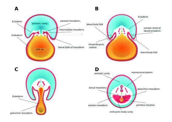
gastroschisis is a congenital structural defect of the abdominal wall, most often to the right of the umbilicus, through which the abdominal viscera protrude. Its developmental, etiological and epidemiological aspects have been a hot topic of controversy for a long time. However, recent findings suggest the involving of genetic and chromosomal alterations and the existence of a stress-inducing pathogenetic pathway, in which risk factors such as demographic and environmental ones can converge. To expand the frontier of knowledge about a malformation that has showed a growing global prevalence, we have conducted a review of the medical literature that gathers information on the embryonic development of the ventral body wall, the primitive intestine, and the ring-umbilical cord complex, as well as on the theories about its origin, pathogenesis and recent epidemiological evidence, for which we consulted bibliographic databases and standard search engines.
La gastrosquisis es un defecto estructural congénito de la pared abdominal, localizado con mayor frecuencia a la derecha del ombligo, a través del cual sobresalen las vísceras abdominales. Durante mucho tiempo, sus aspectos evolutivos, etiológicos y epidemiológicos han sido un tema candente de controversia, aunque hallazgos recientes sugieren la participación de alteraciones genéticas, cromosómicas, y la existencia de una vía patogénica inductora de estrés, en la que factores de riesgo como los demográficos y ambientales pueden converger.Con el objetivo de ampliar la frontera del conocimiento sobre una malformación que ha mostrado una creciente prevalencia global, hemos efectuado una revisión que incluye información, del desarrollo embrionario de la pared corporal ventral, el intestino primitivo, el complejo anillo-cordón umbilical, y de las teorías acerca de su origen, patogénesis e información epidemiológica reciente.
Keywords: Gastroschisis; Hernia umbilical; Vitelline duct; abdominal abnormalities; ectopia cordis; genetic predisposition to disease.
Copyright © 2021 Colombia Medica.
Conflict of interest statement
Conflict of interest: The author do not declare any conflict of interest
Figures
Similar articles
-
Teratogens inducing congenital abdominal wall defects in animal models.Pediatr Surg Int. 2010 Feb;26(2):127-39. doi: 10.1007/s00383-009-2482-z. Epub 2009 Sep 16. Pediatr Surg Int. 2010. PMID: 19756655 Review.
-
Escape of the yolk sac: a hypothesis to explain the embryogenesis of gastroschisis.Clin Genet. 2009 Apr;75(4):326-33. doi: 10.1111/j.1399-0004.2008.01142.x. Clin Genet. 2009. PMID: 19419415
-
Insights into the etiology and embryology of gastroschisis.Semin Pediatr Surg. 2018 Oct;27(5):283-288. doi: 10.1053/j.sempedsurg.2018.08.005. Epub 2018 Aug 27. Semin Pediatr Surg. 2018. PMID: 30413258 Review.
-
The embryologic origin of ventral body wall defects.Semin Pediatr Surg. 2010 Aug;19(3):209-14. doi: 10.1053/j.sempedsurg.2010.03.006. Semin Pediatr Surg. 2010. PMID: 20610194 Review.
-
Anatomy and embryology of abdominal wall defects.Semin Pediatr Surg. 2022 Dec;31(6):151230. doi: 10.1016/j.sempedsurg.2022.151230. Epub 2022 Nov 16. Semin Pediatr Surg. 2022. PMID: 36446303 Review.
Cited by
-
Prenatal cannabis use disorder and gastroschisis in California, 2007-19.Int J Epidemiol. 2024 Feb 14;53(2):dyae042. doi: 10.1093/ije/dyae042. Int J Epidemiol. 2024. PMID: 38503548 Free PMC article.
-
Emerging trends and cross-country health inequalities in congenital birth defects: insights from the GBD 2021 study.Int J Equity Health. 2025 Feb 20;24(1):50. doi: 10.1186/s12939-025-02412-7. Int J Equity Health. 2025. PMID: 39979921 Free PMC article.
-
County-Level Atrazine Use and Gastroschisis.JAMA Netw Open. 2024 May 1;7(5):e2410056. doi: 10.1001/jamanetworkopen.2024.10056. JAMA Netw Open. 2024. PMID: 38709530 Free PMC article.
-
Descriptive epidemiology of gastroschisis in China from 2007 to 2020: a nationwide surveillance-based study.BMC Pediatr. 2024 Sep 14;24(1):584. doi: 10.1186/s12887-024-05056-8. BMC Pediatr. 2024. PMID: 39277760 Free PMC article.
-
Pentalogy of Cantrell Marked With Ectopia Cordis, Gastroschisis, and Cystic Hygroma in the First Trimester: A Rare Case.Cureus. 2024 Sep 13;16(9):e69360. doi: 10.7759/cureus.69360. eCollection 2024 Sep. Cureus. 2024. PMID: 39416577 Free PMC article.
References
-
- Calder J. Two examples of children with preternatural conformation of the guts. Medical Essays and observations. T, W Ruddimans, Medical Society of Edinburgh; 1733.
-
- Torres US, Portela-Oliveira E. Braga F del C.Werner H Jr.Daltro PA.Souza AS When closure fails what the radiologist needs to know about the embryology, anatomy, and prenatal imaging of ventral body wall defects. Semin Ultrasound CT MR. 2015;36(6):522–536. doi: 10.1053/j.sult.2015.01.001. - DOI - PubMed
Publication types
MeSH terms
LinkOut - more resources
Full Text Sources