

COVID-CXNet: Detecting COVID-19 in frontal chest X-ray images using deep learning
- PMID: 35431611
- PMCID: PMC8989406
- DOI: 10.1007/s11042-022-12156-z
COVID-CXNet: Detecting COVID-19 in frontal chest X-ray images using deep learning
Abstract
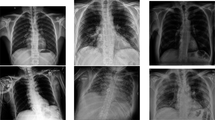
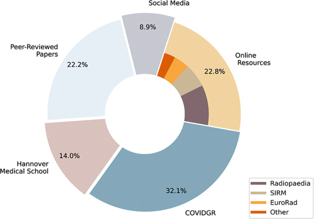
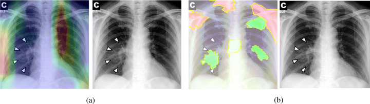
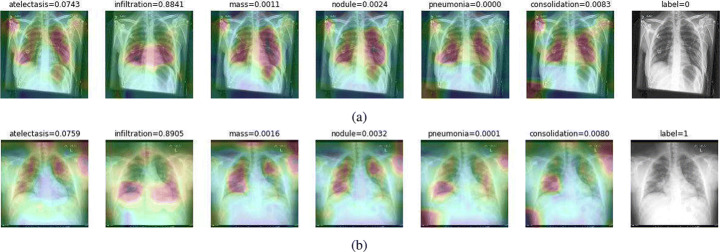
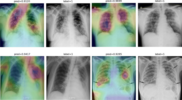
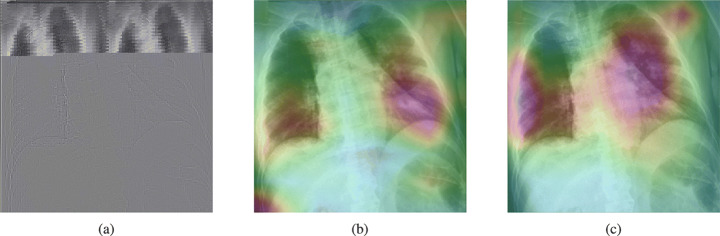
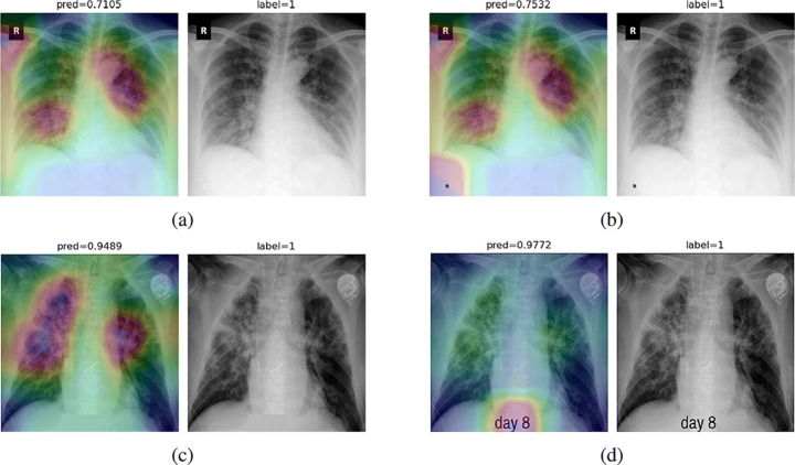
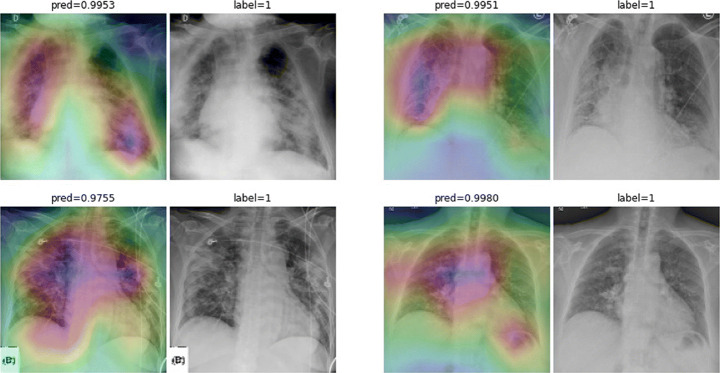
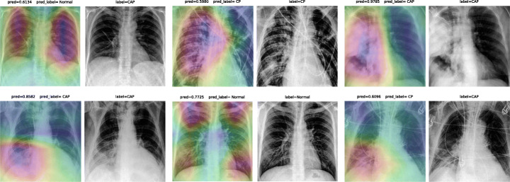
One of the primary clinical observations for screening the novel coronavirus is capturing a chest x-ray image. In most patients, a chest x-ray contains abnormalities, such as consolidation, resulting from COVID-19 viral pneumonia. In this study, research is conducted on efficiently detecting imaging features of this type of pneumonia using deep convolutional neural networks in a large dataset. It is demonstrated that simple models, alongside the majority of pretrained networks in the literature, focus on irrelevant features for decision-making. In this paper, numerous chest x-ray images from several sources are collected, and one of the largest publicly accessible datasets is prepared. Finally, using the transfer learning paradigm, the well-known CheXNet model is utilized to develop COVID-CXNet. This powerful model is capable of detecting the novel coronavirus pneumonia based on relevant and meaningful features with precise localization. COVID-CXNet is a step towards a fully automated and robust COVID-19 detection system.
Keywords: COVID-19; CheXNet; Chest X-ray; Convolutional neural networks; Imaging features.
© The Author(s), under exclusive licence to Springer Science+Business Media, LLC, part of Springer Nature 2022.
Figures

Similar articles
-
Deep Learning Algorithm for COVID-19 Classification Using Chest X-Ray Images.Comput Math Methods Med. 2021 Nov 9;2021:9269173. doi: 10.1155/2021/9269173. eCollection 2021. Comput Math Methods Med. 2021. PMID: 34795794 Free PMC article.
-
CovXNet: A multi-dilation convolutional neural network for automatic COVID-19 and other pneumonia detection from chest X-ray images with transferable multi-receptive feature optimization.Comput Biol Med. 2020 Jul;122:103869. doi: 10.1016/j.compbiomed.2020.103869. Epub 2020 Jun 20. Comput Biol Med. 2020. PMID: 32658740 Free PMC article.
-
COVID-19 Pneumonia Diagnosis Using a Simple 2D Deep Learning Framework With a Single Chest CT Image: Model Development and Validation.J Med Internet Res. 2020 Jun 29;22(6):e19569. doi: 10.2196/19569. J Med Internet Res. 2020. PMID: 32568730 Free PMC article.
-
Detection of coronavirus disease from X-ray images using deep learning and transfer learning algorithms.J Xray Sci Technol. 2020;28(5):841-850. doi: 10.3233/XST-200720. J Xray Sci Technol. 2020. PMID: 32804113 Free PMC article.
-
COVID19XrayNet: A Two-Step Transfer Learning Model for the COVID-19 Detecting Problem Based on a Limited Number of Chest X-Ray Images.Interdiscip Sci. 2020 Dec;12(4):555-565. doi: 10.1007/s12539-020-00393-5. Epub 2020 Sep 21. Interdiscip Sci. 2020. PMID: 32959234 Free PMC article.
Cited by
-
Survey of Explainable AI Techniques in Healthcare.Sensors (Basel). 2023 Jan 5;23(2):634. doi: 10.3390/s23020634. Sensors (Basel). 2023. PMID: 36679430 Free PMC article. Review.
-
Machine-Learning-Based Disease Diagnosis: A Comprehensive Review.Healthcare (Basel). 2022 Mar 15;10(3):541. doi: 10.3390/healthcare10030541. Healthcare (Basel). 2022. PMID: 35327018 Free PMC article. Review.
-
Deep Learning for Reliable Classification of COVID-19, MERS, and SARS from Chest X-ray Images.Cognit Comput. 2022;14(5):1752-1772. doi: 10.1007/s12559-021-09955-1. Epub 2022 Jan 11. Cognit Comput. 2022. PMID: 35035591 Free PMC article.
-
Validation of expert system enhanced deep learning algorithm for automated screening for COVID-Pneumonia on chest X-rays.Sci Rep. 2021 Dec 1;11(1):23210. doi: 10.1038/s41598-021-02003-w. Sci Rep. 2021. PMID: 34853342 Free PMC article.
-
Chest X-ray image phase features for improved diagnosis of COVID-19 using convolutional neural network.Int J Comput Assist Radiol Surg. 2021 Feb;16(2):197-206. doi: 10.1007/s11548-020-02305-w. Epub 2021 Jan 9. Int J Comput Assist Radiol Surg. 2021. PMID: 33420641 Free PMC article.
References
-
- Al-Karawi D, Al-Zaidi S, Polus N, Jassim S (2020) Ai based chest x-ray (cxr) scan texture analysis algorithm for digital test of covid-19 patients. medRxiv
-
- Almuhayar M, Lu HH-S, Iriawan N (2019) Classification of abnormality in chest x-ray images by transfer learning of chexnet. In: 2019 3Rd international conference on informatics and computational sciences (ICICos). IEEE, pp 1–6
-
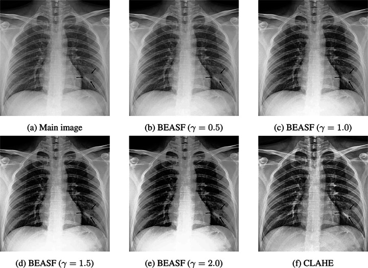
- Arriaga-Garcia EF, Sanchez-Yanez RE, Garcia-Hernandez M (2014) Image enhancement using bi-histogram equalization with adaptive sigmoid functions. In: 2014 International conference on electronics, communications and computers (CONIELECOMP). IEEE, pp 28–34
LinkOut - more resources
Full Text Sources