

Anti-Epileptic Effect of Crocin on Experimental Temporal Lobe Epilepsy in Mice
- PMID: 35431921
- PMCID: PMC9009530
- DOI: 10.3389/fphar.2022.757729
Anti-Epileptic Effect of Crocin on Experimental Temporal Lobe Epilepsy in Mice
Abstract
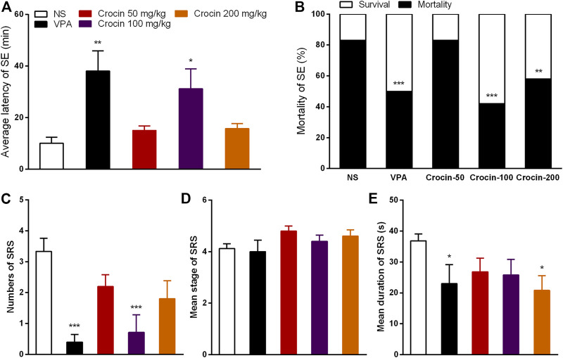
Temporal lobe epilepsy (TLE) is a common kind of refractory epilepsy. More than 30% TLE patients were multi-drug resistant. Some patients may even develop into status epilepticus (SE) because of failing to control seizures. Thus, one of the avid goals for anti-epileptic drug development is to discover novel potential compounds to treat TLE or even SE. Crocin, an effective component of Crocus sativus L., has been applied in several epileptogenic models to test its anti-epileptic effect. However, it is still controversial and its effect on TLE remains unclear. Therefore, we investigated the effects of crocin on epileptogenesis, generalized seizures (GS) in hippocampal rapid electrical kindling model as well as SE and spotaneous recurrent seizure (SRS) in pilocarpine-induced TLE model in ICR mice in this study. The results showed that seizure stages and cumulative afterdischarge duration were significantly depressed by crocin (20 and 50 mg/kg) during hippocampal rapid kindling acquisition. And crocin (100 mg/kg) significantly reduced the incidence of GS and average seizure stages in fully kindled animals. In pilocarpine-induced TLE model, the latency of SE was significantly prolonged and the mortality of SE was significantly decreased by crocin (100 mg/kg), which can also significantly suppress the number of SRS. The underlying mechanism of crocin may be involved in the protection of neurons, the decrease of tumor necrosis factor-α in the hippocampus and the increase of brain derived neurotrophic factor in the cortex. In conclusion, crocin may be a potential and promising anti-epileptic compound for treatment of TLE.
Keywords: crocin; kindling; pilocarpine; spontaneous recurrent seizure; temporal lobe epilepsy.
Copyright © 2022 Zhong, Qian, Lyu, Wang, Hu, Yu, Ma and Ye.
Conflict of interest statement
The authors declare that the research was conducted in the absence of any commercial or financial relationships that could be construed as a potential conflict of interest.
Figures


Similar articles
-
[Effects of crocin on hippocampus rapid kindling epilepsy in mice].Zhejiang Da Xue Xue Bao Yi Xue Ban. 2017 Jan 25;46(1):7-14. doi: 10.3785/j.issn.1008-9292.2017.02.02. Zhejiang Da Xue Xue Bao Yi Xue Ban. 2017. PMID: 28436625 Free PMC article. Chinese.
-
Soluble epoxide hydrolase activity regulates inflammatory responses and seizure generation in two mouse models of temporal lobe epilepsy.Brain Behav Immun. 2015 Jan;43:118-29. doi: 10.1016/j.bbi.2014.07.016. Epub 2014 Aug 15. Brain Behav Immun. 2015. PMID: 25135858
-
Behavioral and histological assessment of the effect of intermittent feeding in the pilocarpine model of temporal lobe epilepsy.Epilepsy Res. 2009 Sep;86(1):54-65. doi: 10.1016/j.eplepsyres.2009.05.003. Epub 2009 Jun 7. Epilepsy Res. 2009. PMID: 19505798
-
Animal models of epilepsy for the development of antiepileptogenic and disease-modifying drugs. A comparison of the pharmacology of kindling and post-status epilepticus models of temporal lobe epilepsy.Epilepsy Res. 2002 Jun;50(1-2):105-23. doi: 10.1016/s0920-1211(02)00073-6. Epilepsy Res. 2002. PMID: 12151122 Review.
-
Molecular neuropathology of temporal lobe epilepsy: complementary approaches in animal models and human disease tissue.Epilepsia. 2007;48 Suppl 2:4-12. doi: 10.1111/j.1528-1167.2007.01062.x. Epilepsia. 2007. PMID: 17571348 Review.
Cited by
-
Advances on the anti-tumor mechanisms of the carotenoid Crocin.PeerJ. 2023 Jun 29;11:e15535. doi: 10.7717/peerj.15535. eCollection 2023. PeerJ. 2023. PMID: 37404473 Free PMC article. Review.
-
Oral administration of crocin-loaded solid lipid nanoparticles inhibits neuroinflammation in a rat model of epileptic seizures by activating SIRT1 expression.Res Pharm Sci. 2024 Aug 16;19(4):397-414. doi: 10.4103/RPS.RPS_68_24. eCollection 2024 Aug. Res Pharm Sci. 2024. PMID: 39399725 Free PMC article.
-
Establishing a Mouse Model of NL3R617W-Associated Autism Spectrum Disorder for a Functional Study.Actas Esp Psiquiatr. 2025 Mar;53(2):253-266. doi: 10.62641/aep.v53i2.1780. Actas Esp Psiquiatr. 2025. PMID: 40071366 Free PMC article.
-
Research progress on the pharmacological activity, biosynthetic pathways, and biosynthesis of crocins.Beilstein J Org Chem. 2024 Apr 9;20:741-752. doi: 10.3762/bjoc.20.68. eCollection 2024. Beilstein J Org Chem. 2024. PMID: 38633914 Free PMC article. Review.
-
Protective effect of crocin-loaded nanoparticles in rats following epileptic seizures: Biochemical, behavioral and histopathological outcomes.Heliyon. 2024 Aug 10;10(16):e36122. doi: 10.1016/j.heliyon.2024.e36122. eCollection 2024 Aug 30. Heliyon. 2024. PMID: 39229546 Free PMC article.
References
LinkOut - more resources
Full Text Sources