. 2022 Apr 1:13:832611.
doi: 10.3389/fphar.2022.832611. eCollection 2022.

Acute Administration of Metformin Protects Against Neuronal Apoptosis Induced by Cerebral Ischemia-Reperfusion Injury via Regulation of the AMPK/CREB/BDNF Pathway

Affiliations

Acute Administration of Metformin Protects Against Neuronal Apoptosis Induced by Cerebral Ischemia-Reperfusion Injury via Regulation of the AMPK/CREB/BDNF Pathway

Ke Liu et al. Front Pharmacol. .

Erratum in

Abstract

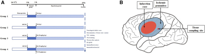
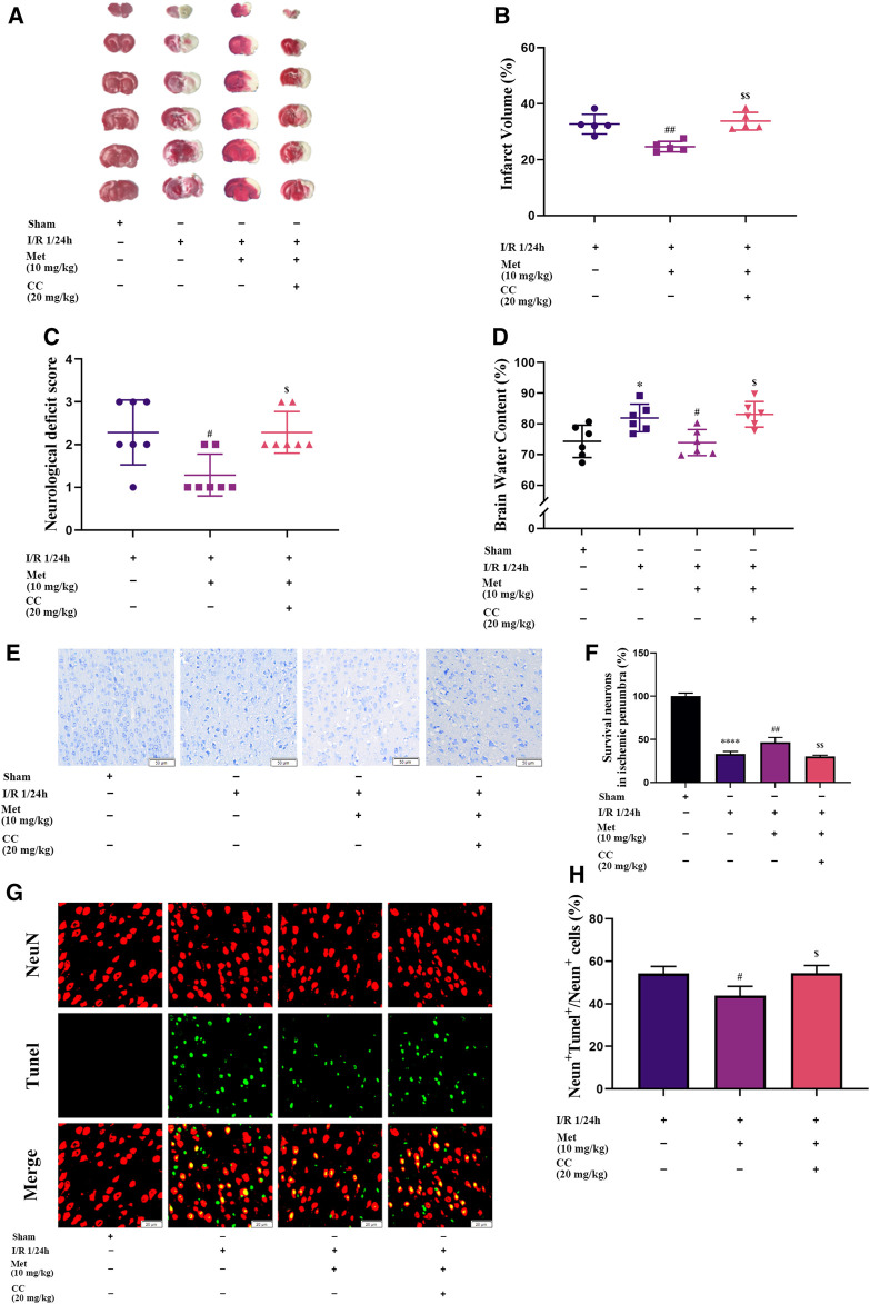
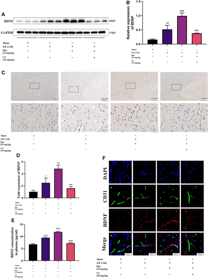
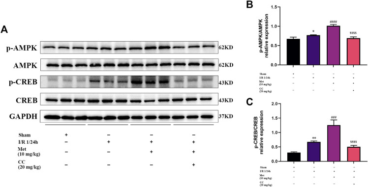
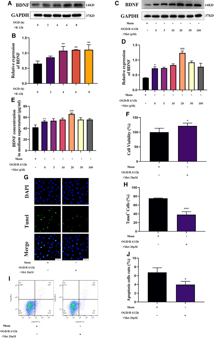
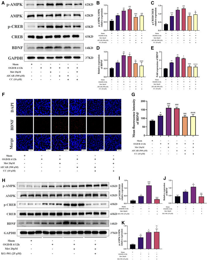
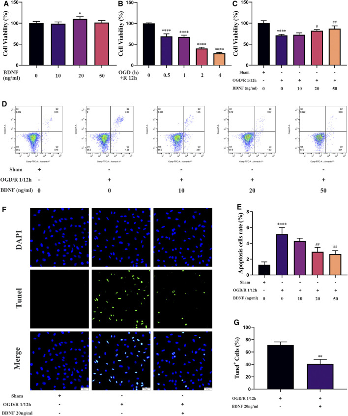
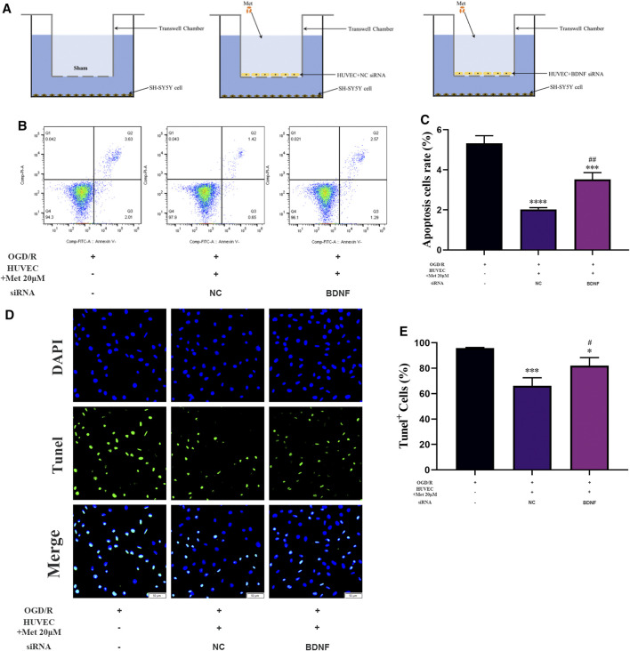
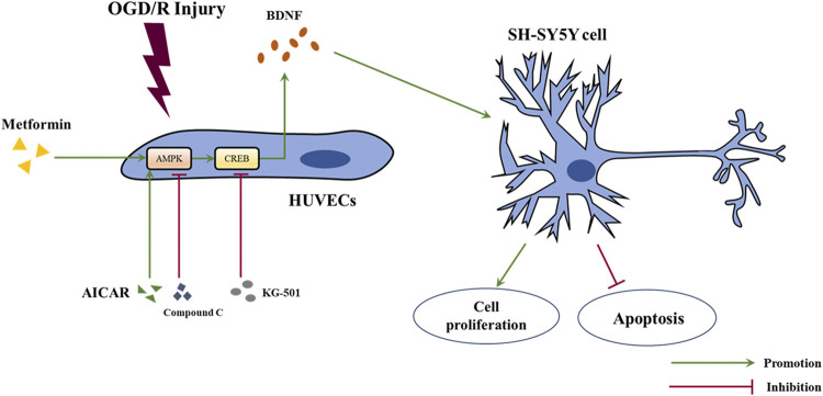
Metformin is a first-line anti-diabetic agent with a powerful hypoglycemic effect. Several studies have reported that metformin can improve the prognosis of stroke patients and that this effect is independent of its hypoglycemic effect; however, the specific mechanism remains unclear. In this research, we explored the effect and specific mechanism of metformin in cerebral ischemia-reperfusion (I/R) injury by constructing a transient middle cerebral artery occlusion model in vivo and a glucose and oxygen deprivation/reoxygenation (OGD/R) model in vitro. The results of the in vivo experiments showed that acute treatment with low-dose metformin (10 mg/kg) ameliorated cerebral edema, reduced the cerebral infarction volume, improved the neurological deficit score, and ameliorated neuronal apoptosis in the ischemic penumbra. Moreover, metformin up-regulated the brain-derived neurotrophic factor (BDNF) expression and increased phosphorylation levels of AMP-activated protein kinase (AMPK) and cAMP-response element binding protein (CREB) in the ischemia penumbra. Nevertheless, the above-mentioned effects of metformin were reversed by Compound C. The results of the in vitro experiments showed that low metformin concentrations (20 μM) could reduce apoptosis of human umbilical vein endothelial cells (HUVECs) under OGD/R conditions and promote cell proliferation. Moreover, metformin could further promote BDNF expression and release in HUVECs under OGD/R conditions via the AMPK/CREB pathway. The Transwell chamber assay showed that HUVECs treated with metformin could reduce apoptosis of SH-SY5Y cells under OGD/R conditions and this effect could be partially reversed by transfection of BDNF siRNA in HUVECs. In summary, our results suggest that metformin upregulates the level of BDNF in the cerebral ischemic penumbra via the AMPK/CREB pathway, thereby playing a protective effect in cerebral I/R injury.

Keywords: AMP-activated protein kinase; brain-derived neurotrophic factor; cerebral ischemia-reperfusion injury; metformin; neuronal apoptosis.


Conflict of interest statement

The authors declare that the research was conducted in the absence of any commercial or financial relationships that could be construed as a potential conflict of interest.

Figures

FIGURE 1
FIGURE 1
Schematic diagram of animal experiment groupings and ischemic penumbra. (A) The schematic diagram of animal experiment groupings (n = 17 in each group). Abbreviations: tMCAO, transient middle cerebral artery occlusion; Veh, vehicle; Met, metformin; CC, Compound C. (B) The schematic diagram of ischemic penumbra.
FIGURE 2
FIGURE 2
Metformin alleviates cerebral edema, reduces cerebral infarct volume, improves the neurological deficit score, and ameliorates neuronal apoptosis in the ischemic penumbra. However, Compound C significantly reduces the above-mentioned effects of metformin. (A,B) Cerebral infarct volume was assessed by TTC staining (n = 5). (C) The neurological deficit score was assessed by the Zea-Longa five-point scoring system (n = 7). (D) Brain edema was evaluated by measuring the brain water content (n = 6). (E–H) Survival and apoptosis of neurons around the ischemic penumbra were evaluated by Nissl staining (n = 3, bar = 50 μm) and TUNEL staining (n = 3, bar = 20 μm), respectively. (*p < 0.05, ****p < 0.0001 vs. sham; # p < 0.05, ## p < 0.01 vs. I/R 1/24 h; $ p < 0.05, $$ p < 0.01, vs. I/R 1/24 h + Met).
FIGURE 3
FIGURE 3
Metformin improves cerebral ischemia/reperfusion (I/R) injury by regulating the expression of BDNF. (A–C) BDNF protein expression in the ischemic penumbra area was evaluated by western blotting and immunohistochemistry (n = 3, bar = 100 μm/20 μm). (D) The BDNF transcription level in the ischemic penumbra area was assessed by RT-qPCR (n = 3). (E) The BDNF concentration in the plasma assessed by ELISA (n = 3). (F) BDNF expression in endothelial cells in the ischemic penumbra was evaluated by immunofluorescence staining (n = 3, bar = 20 μm). (*p < 0.05, **p < 0.01, ****p < 0.0001 vs. sham; ## p < 0.01, ### p < 0.001, #### p < 0.0001 vs. I/R 1/24 h; $$$ p < 0.001, $$$$ p < 0.0001 vs. I/R 1/24 h + Met).
FIGURE 4
FIGURE 4
AMPK and CREB are involved in the regulation of BDNF expression by metformin in cerebral ischemia-reperfusion (I/R) injury. (A–C) The phosphorylation levels of AMPK and CREB proteins was detected by western blotting (n = 3). (* p < 0.05, ** p < 0.01 vs. sham; ### p < 0.001, #### p < 0.0001 vs. I/R 1/24 h; $$$$ p < 0.0001 vs. I/R 1/24 h + Met).
FIGURE 5
FIGURE 5
Metformin upregulates BDNF expression and release, promotes HUVECs proliferation, and inhibits cell apoptosis under OGD/R conditions. (A–D) The BDNF expression level of metformin-treated HUVECs was detected by western blotting (n = 3). (E) The BDNF concentration in the culture supernatant of metformin-treated HUVECs was detected by ELISA (n = 3). (F) Proliferation of HUVECs was detected by CCK-8 assay (n = 5). (G–J) Apoptosis of HUVECs was detected by TUNEL staining (n = 3, bar = 50 μm) and flow cytometry (n = 3). (* p < 0.05, ** p < 0.01 vs. sham; # p < 0.05, ### p < 0.001 vs. OGD/R 6/12 h).
FIGURE 6
FIGURE 6
Metformin promotes the expression of BDNF in HUVECs under OGD/R conditions through the AMPK/CREB pathway. (A–D) The BDNF expression level and AMPK and CREB phosphorylation levels in HUVECs treated with metformin, AICAR, and Compound C under OGD/R conditions detected were by western blotting (n = 3). (E) The BDNF transcription level under the above conditions was detected by RT-qPCR (n = 4). (F,G) Immunofluorescence staining and quantification of BDNF under the above conditions (n = 3, bar = 20 μM). (H–K) BDNF expression level and AMPK and CREB phosphorylation levels in HUVECs treated with metformin and KG-501 under OGD/R conditions were detected by western blotting (n = 3). (* p < 0.05, ** p < 0.01, *** p < 0.001 vs. sham; # p < 0.05, ## p < 0.01, ### p < 0.001, #### p < 0.0001 vs. OGD/R 6/12 h; NS, not significant, $$ p < 0.01, $$$ p < 0.001, $$$$ p < 0.0001 vs. OGD/R 6/12 h + AICAR; & p < 0.05, && p < 0.01, &&& p < 0.001, &&&& p < 0.0001 vs. OGD/R 6/12 h + Met 20 μM).
FIGURE 7
FIGURE 7
BDNF promotes the proliferation of SH-SY5Y cells and inhibits cell apoptosis under OGD/R conditions. (A–C) The effect of BDNF on the proliferation of SH-SY5Y cells was detected by CCK-8 assay under OGD/R conditions (n = 4). (D–G) The effect of BDNF on apoptosis of SH-SY5Y cells was detected by TUNEL staining (n = 3, bar = 50 μm) and flow cytometry (n = 3). (* p < 0.05, ** p < 0.01, **** p < 0.0001 vs. sham; # p < 0.05, ## p < 0.01 vs. OGD/R 1/12 h).
FIGURE 8
FIGURE 8
HUVECs treated with metformin reduce apoptosis of SH-SY5Y cells under OGD/R conditions. (A) Schematic diagram of grouping in the Transwell chamber assay. (B–E) Apoptosis of SH-SY5Y cells was detected by TUNEL staining (n = 3, bar = 50 μm) and flow cytometry (n = 3). (* p < 0.05, *** p < 0.001, **** p < 0.0001 vs. OGD/R; # p < 0.05, ## p < 0.01 vs. OGD/R + HUVEC + Met 20 μM + NC siRNA; NC, negative control).
FIGURE 9
FIGURE 9
Schematic diagram of the mechanism of metformin in this study. Abbreviations: OGD/R, oxygen-glucose deprivation/reoxygenation; HUVECs, human umbilical vein endothelial cells; BDNF, brain-derived neurotrophic factor.

Similar articles

Cited by

References

    1. Alexander S. P., Benson H. E., Faccenda E., Pawson A. J., Sharman J. L., Spedding M., et al. (2013). The Concise Guide to PHARMACOLOGY 2013/14: Enzymes. Br. J. Pharmacol. 170 (8), 1797–1867. 10.1111/bph.12451 - DOI - PMC - PubMed
    1. Alhusban A., Kozak A., Ergul A., Fagan S. C. (2013). AT1 Receptor Antagonism Is Proangiogenic in the Brain: BDNF a Novel Mediator. J. Pharmacol. Exp. Ther. 344 (2), 348–359. 10.1124/jpet.112.197483 - DOI - PMC - PubMed
    1. Ashabi G., Khodagholi F., Khalaj L., Goudarzvand M., Nasiri M. (2014). Activation of AMP-Activated Protein Kinase by Metformin Protects against Global Cerebral Ischemia in Male Rats: Interference of AMPK/PGC-1α Pathway. Metab. Brain Dis. 29 (1), 47–58. 10.1007/s11011-013-9475-2 - DOI - PubMed
    1. Ashabi G., Khalaj L., Khodagholi F., Goudarzvand M., Sarkaki A. (2015). Pre-treatment with Metformin Activates Nrf2 Antioxidant Pathways and Inhibits Inflammatory Responses through Induction of AMPK after Transient Global Cerebral Ischemia. Metab. Brain Dis. 30 (3), 747–754. 10.1007/s11011-014-9632-2 - DOI - PubMed
    1. Bekinschtein P., Cammarota M., Medina J. H. (2014). BDNF and Memory Processing. Neuropharmacology 76, 677–683. 10.1016/j.neuropharm.2013.04.024 - DOI - PubMed

LinkOut - more resources