. 2022 Apr 6:2022:5402997.
doi: 10.1155/2022/5402997. eCollection 2022.

Selenium Supplementation Improved Cardiac Functions by Suppressing DNMT2-Mediated GPX1 Promoter DNA Methylation in AGE-Induced Heart Failure

Affiliations

Selenium Supplementation Improved Cardiac Functions by Suppressing DNMT2-Mediated GPX1 Promoter DNA Methylation in AGE-Induced Heart Failure

Huolan Zhu et al. Oxid Med Cell Longev. .

Abstract

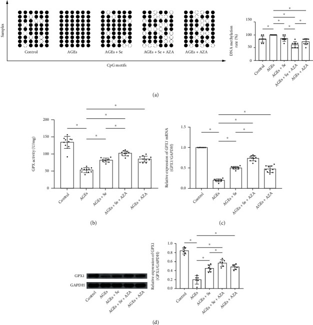
Objective: Advanced glycation end products (AGEs) are featured metabolites associated with diabetic cardiomyopathy which is characterized by heart failure caused by myocyte apoptosis. Selenium was proved cardioprotective. This study was aimed at investigating the therapeutic effects and underlying mechanisms of selenium supplementation on AGE-induced heart failure.

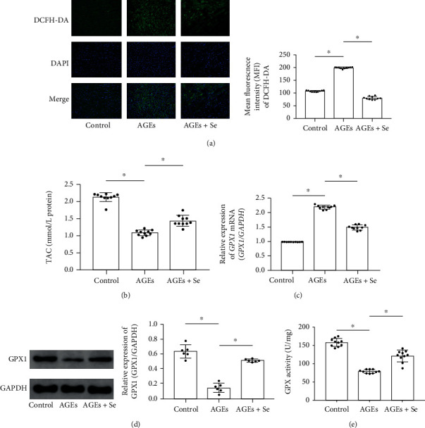
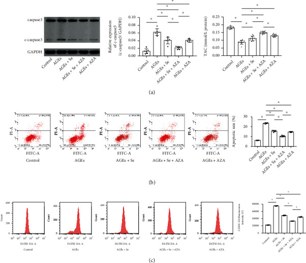
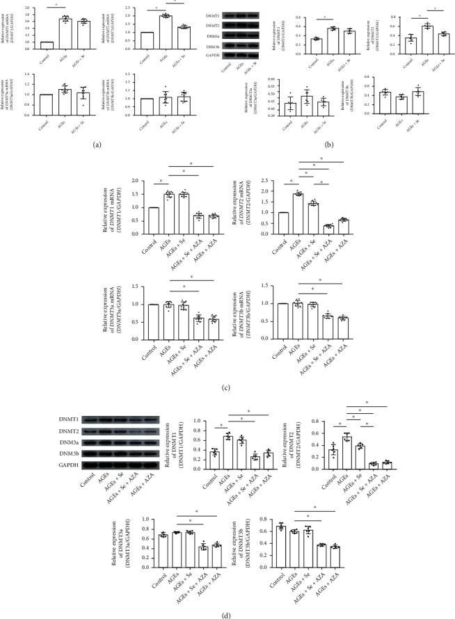
Methods: Rats and primary myocytes were exposed to AGEs. Selenium supplementation was administrated. Cardiac functions and myocyte apoptosis were evaluated. Oxidative stress was assessed by total antioxidant capacity (TAC), reactive oxygen species (ROS) generation, and GPX activity. Expression levels of DNA methyltransferases (DNMTs) and glutathione peroxidase 1 (GPX1) were evaluated. DNA methylation of the GPX1 promoter was analyzed.

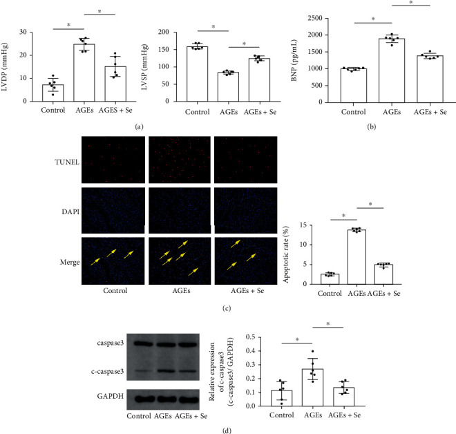
Results: AGE exposure elevated intracellular ROS generation, induced myocyte apoptosis, and impaired cardiac functions. AGE exposure increased DNMT1 and DNMT2 expression, leading to the reduction of GPX1 expression and activity in the heart. Selenium supplementation decreased DNMT2 expression, recovered GPX1 expression and activity, and alleviated intracellular ROS generation and myocyte apoptosis, resulting in cardiac function recovery. DNA methylation analysis in primary myocytes indicated that selenium supplementation or DNMT inhibitor AZA treatment reduced DNA methylation of the GPX1 gene promoter. Selenium supplementation and AZA administration showed synergic inhibitory effect on GPX1 gene promoter methylation.

Conclusions: Selenium supplementation showed cardioprotective effects on AGE-induced heart failure by suppressing ROS-mediated myocyte apoptosis. Selenium supplementation suppressed ROS generation by increasing GPX1 expression via inhibiting DNMT2-induced GPX1 gene promoter DNA methylation in myocytes exposed to AGEs.


Conflict of interest statement

All authors declare no conflicts of interest.

Figures

Figure 1
Figure 1
(a) Columns indicated the calculated left ventricular systolic pressure (LVSP) and left ventricular diastolic pressure (LVDP) in rats in control, AGE, and AGE + Se groups. (b) Columns indicated the detected BNP concentrations in blood samples collected from rats in control, AGE, and AGE + Se groups. (c) Captured fluorescent images of TUNEL assay of cardiac tissue. TUNEL-positive cells were tagged with red fluorescence and pointed by yellow arrows. Columns indicated the apoptosis rate in cardiac tissue harvested from rats in control, AGE, and AGE + Se groups, respectively. (d) Immunoblots of caspase3, cleaved caspase3 (c-caspase3), and GAPDH were demonstrated. Columns indicated relative expression levels of c-caspase3 in cardiac tissue harvested from rats in control, AGE, and AGE + Se groups, respectively. Magnification of images in Figure 1(a) was 200 (control: rats treated with control BSA; AGEs: rats treated with AGE-BSA; AGEs + Se: rats treated with AGE-BSA then supplemented with sodium selenite (n = 6, P < 0.05)).
Figure 2
Figure 2
(a) Images of ROS indicator DCFH-DA stains of cardiac tissue sections were demonstrated. Columns indicated mean fluorescent intensities of DCFH-DA in control, AGE, AGE + Se groups, respectively. (b) Columns indicated the detected total antioxidant capacity (TAC) of cardiac tissue homogenates from rats in control, AGE, and AGE + Se groups. (c) Columns indicated relative expression levels of GPX1 mRNA in cardiac tissue of rats from control, AGE, and AGE + Se groups. (d) Immunoblots of GPX1 and GAPDH were demonstrated. Columns indicated relative expression of GPX1. (e) Columns indicated measured GPX activity cardiac tissue homogenates from rats in control, AGE, and AGE + Se groups. Magnification of images in Figure 2(a) was 200 (control: rats treated with control BSA; AGEs: rats treated with AGE-BSA; AGEs + Se: rats treated with AGE-BSA then supplemented with sodium selenite (n = 10, P < 0.05)).
Figure 3
Figure 3
(a) Immunoblots of caspase3, cleaved caspase3 (c-caspase3), and GAPDH were demonstrated. Columns indicated relative expression levels of c-caspase3 in myocytes from control, AGEs, AGE + Se, AGE + Se + AZA, and AGE + AZA groups. (b) Plotted charts of flow cytometry detecting apoptotic myocytes were demonstrated. Columns indicated the apoptotic rate of myocytes from control, AGE, AGE + Se, AGE + Se + AZA, and AGE + AZA groups. (c) Plotted charts of flow cytometry measuring DCFH-DA fluorescence in myocytes were demonstrated. Columns indicated fluorescent intensities of DCFH-DA in myocytes from control, AGE, AGE + Se, AGE + Se + AZA, and AGE + AZA groups (control: myocytes treated with control BSA; AGEs: myocytes incubated with AGEs; AGEs + Se: AGE-exposed myocytes treated with sodium selenite; AGEs + Se + AZA: AGE-exposed myocytes treated with sodium selenite and AZA; AGEs + AZA: AGE-exposed myocytes treated with AZA (n = 10, P < 0.05)).
Figure 4
Figure 4
(a) Columns indicated relative mRNA expression levels of DNMT1, DNMT2, DNMT3a, and DNMT3b in cardiac tissue of rats from control, AGE, and AGE + Se groups. (b) Immunoblots of DNMT1, DNMT2, DNMT3a, and DNMT3b. Columns indicated relative expression levels of DNMT1, DNMT2, DNMT3a, and DNMT3b in cardiac tissue of rats from control, AGE, and AGE + Se groups (control: rats treated with control BSA; AGEs: rats treated with AGE-BSA; AGEs + Se: rats treated with AGE-BSA then supplemented with sodium selenite (n = 6 or 10, P < 0.05)). (c) Columns indicated relative mRNA expression levels of DNMT1, DNMT2, DNMT3a, and DNMT3b in myocytes from control, AGEs, AGE + Se, AGE + Se + AZA, and AGE + AZA groups, respectively. (d) Immunoblots of DNMT1, DNMT2, DNMT3a, and DNMT3b. Columns indicated relative expression levels of DNMT1, DNMT2, DNMT3a, and DNMT3b in myocytes from control, AGEs, AGE + Se, AGE + Se + AZA, and AGE + AZA groups, respectively (control: myocytes treated with control BSA; AGEs: myocytes incubated with AGEs; AGEs + Se: AGE-exposed myocytes treated with sodium selenite; AGEs + Se + AZA: AGE-exposed myocytes treated with sodium selenite and AZA; AGEs + AZA: AGE-exposed myocytes treated with AZA (n = 6 or 10, P < 0.05)).
Figure 5
Figure 5
(a) Diagram of DNA methylation of the GPX1 gene promoter was plotted. Black dots indicated methylated sites while white dots indicated demethylated sites. Columns indicated the methylation rate of the GPX1 gene promoter in myocytes from control, AGE, AGE + Se, AGEs+Se + AZA, and AGE + AZA groups (control: myocytes treated with control BSA; AGEs: myocytes incubated with AGEs; AGEs + Se: AGE-exposed myocytes treated with sodium selenite; AGEs + Se + AZA: AGE-exposed myocytes treated with sodium selenite and AZA; AGEs + AZA: AGE-exposed myocytes treated with AZA (n = 9, P < 0.05)); (b) columns indicated the GPX activity in myocytes from control, AGE, AGE + Se, AGE + Se + AZA, and AGE + AZA groups. (c) Columns indicated relative expression levels of GPX1 mRNA in myocytes from control, AGEs, AGE + Se, AGE + Se + AZA, and AGE + AZA groups. (d) Immunoblots of GPX1 and GAPDH were demonstrated. Columns indicated relative expression levels of GPX1 in myocytes from control, AGE, AGE + Se, AGE + Se + AZA, and AGE + AZA groups (control: myocytes treated with control BSA; AGEs: myocytes incubated with AGEs; AGEs + Se: AGE-exposed myocytes treated with sodium selenite; AGEs + Se + AZA: AGE-exposed myocytes treated with sodium selenite and AZA; AGEs + AZA: AGE-exposed myocytes treated with AZA (n = 6 or 10, P < 0.05)).

References

    1. Dillmann W. H. Diabetic cardiomyopathy. Circulation Research . 2019;124(8):1160–1162. doi: 10.1161/CIRCRESAHA.118.314665. - DOI - PMC - PubMed
    1. Liu Z., Zhao N., Zhu H., et al. Circulating interleukin-1β promotes endoplasmic reticulum stress-induced myocytes apoptosis in diabetic cardiomyopathy via interleukin-1 receptor-associated kinase-2. Cardiovascular Diabetology . 2015;14(1):p. 125. doi: 10.1186/s12933-015-0288-y. - DOI - PMC - PubMed
    1. Wu W., Liu X., Han L. Apoptosis of cardiomyocytes in diabetic cardiomyopathy involves overexpression of glycogen synthase kinase-3β. Bioscience Reports . 2019;39(1) doi: 10.1042/BSR20171307. - DOI - PMC - PubMed
    1. Jia G., Hill M. A., Sowers J. R. Diabetic cardiomyopathy: an update of mechanisms contributing to this clinical entity. Circulation Research . 2018;122(4):624–638. doi: 10.1161/CIRCRESAHA.117.311586. - DOI - PMC - PubMed
    1. Bodiga V. L., Eda S. R., Bodiga S. Advanced glycation end products: role in pathology of diabetic cardiomyopathy. Heart Failure Reviews . 2014;19(1):49–63. doi: 10.1007/s10741-013-9374-y. - DOI - PubMed