. 2022 Apr 7:2022:4441603.
doi: 10.1155/2022/4441603. eCollection 2022.

Impact of Yiqi Huoxue Decoction on the Relationship between Remodeling of Cardiac Nerves and Macrophages after Myocardial Infarction in Rats

Affiliations

Impact of Yiqi Huoxue Decoction on the Relationship between Remodeling of Cardiac Nerves and Macrophages after Myocardial Infarction in Rats

Yunke Liu et al. J Healthc Eng. .

Retraction in

Abstract

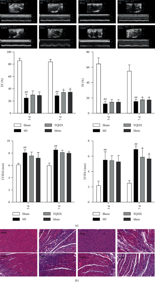
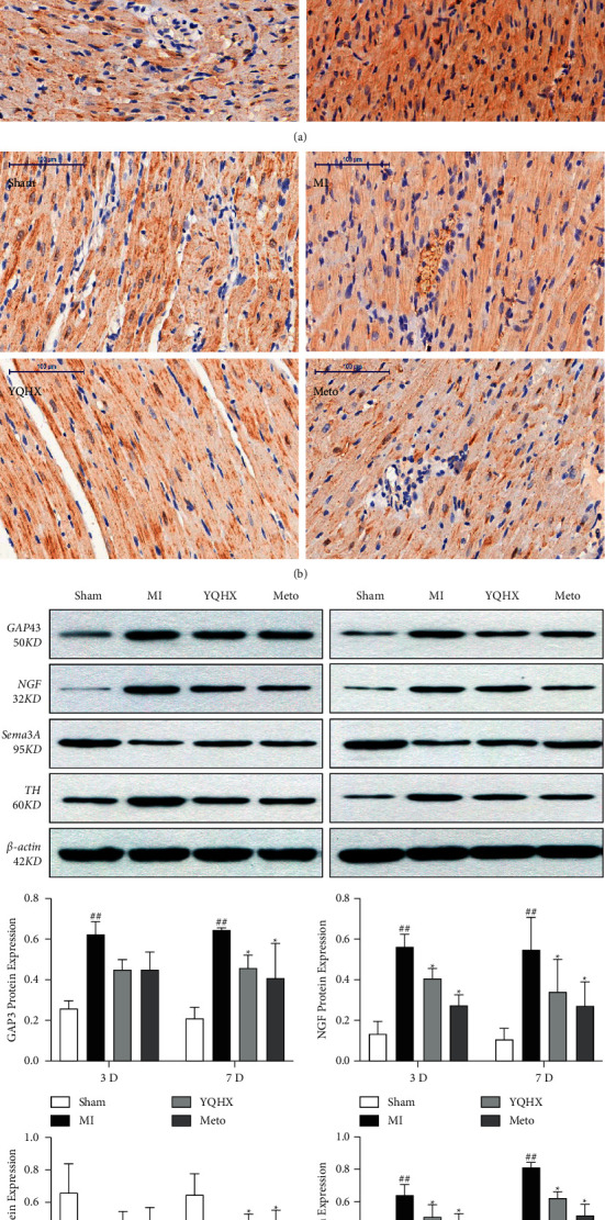
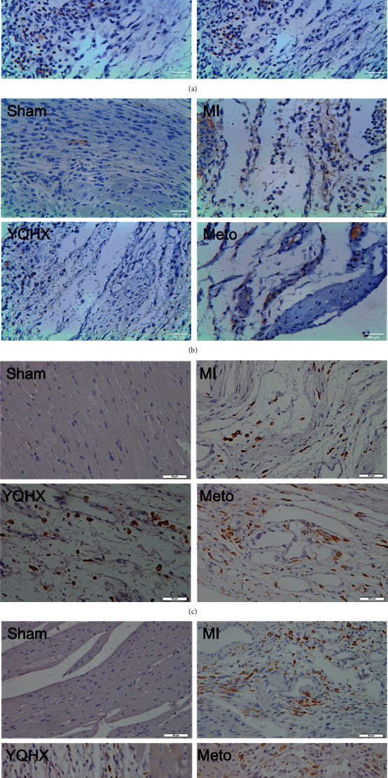
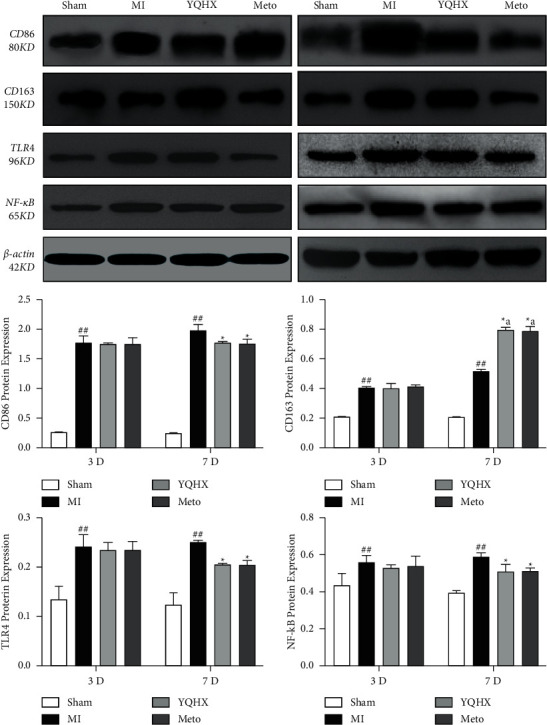
Sympathetic nerve remodeling after myocardial infarction (MI) has an indispensable role in cardiac remodeling. Numerous works have shown that sympathetic nerve remodeling can be delayed by inhibition of inflammatory response. Earlier studies have shown improvement in ventricular remodeling and inhibited chronic stage neural remodeling by Yiqi Huoxue decoction (YQHX). Therefore, the current study looked at the inhibitory effect of YQHX prescription on proinflammatory mediators and macrophages and the effect on neural remodeling at 3 and 7 days after MI. YQHX inhibited the expression of Toll-like receptor 4 (TLR4) and nuclear factor kappa B (NF-κB) proteins and macrophage infiltration within 7 days after myocardial infarction. YQHX could decrease Th-positive nerve fiber density in the area around infarction and reduce the expression of growth-associated protein 43 (GAP43), nerve growth factor (NGF), and tyrosine hydroxylase (TH) proteins, which was associated with the remodeling of sympathetic nerves. Thus, the nerve remodeling inhibition after MI due to YQHX may be through its anti-inflammatory action. These data provide direct evidence for the potential application of traditional Chinese medicine (TCM) in the remodeling of sympathetic nerves after MI.


Conflict of interest statement

The authors declare that they have no conflicts of interest.

Figures

Figure 1
Figure 1
(a) Cardiac function and echocardiogram of rats in each group after days 3 and 7 of MI (myocardial infarction) (N = 9). ##P < 0.01, vs. the sham operation group; P < 0.01, vs. the MI model group. (b) Histopathological staining using hematoxylin and eosin of the tissue in the infarct marginal area of rats with MI in each group (scale bar = 200 μm).
Figure 2
Figure 2
(a) Immunohistochemical staining for examining the positive, sympathetic nerve fibers at the edge of MI (myocardial infarction) in rats 3 days after MI (scale). (b) Immunohistochemical staining was used to observe the positive, sympathetic nerve fibers at the edge of myocardial infarction in rats 7 days after MI (myocardial infarction). (c) The influence of YQHX on GAP43, TH, SEMA-3A, and NGF expression. ##P < 0.01, vs. the sham operation group; P < 0.01, vs. MI.
Figure 3
Figure 3
(a) Immunohistochemical analysis to detect the CD86 macrophages at the edge of myocardial infarction (MI) in rats 3 days after myocardial infarction. (b) Immunohistochemical analysis to observe the CD86 macrophages at the edge of MI in rats 7 days after myocardial infarction. (c) Immunohistochemical analysis to observe the CD163 macrophages at the edge of MI in rats 3 days following myocardial infarction. (d) Immunohistochemical analysis to observe the CD163 macrophages at the edge of MI in rats 7 days after myocardial infarction.
Figure 4
Figure 4
The expression of CD86, CD163, TLR4, and NFκB in each group of rats; the tissue of the infarct marginal area was examined. ##P < 0.01, vs. the sham operation group; P < 0.01, vs. the MI model group; aP < 0.01, vs. the model group of 3 days.

Similar articles

Cited by

References

    1. Jenča D., Melenovský V., Stehlik J., et al. Heart failure after myocardial infarction: incidence and predictors. ESC Heart Failure . 2021 February;8(1):222–237. doi: 10.1002/ehf2.13144. - DOI - PMC - PubMed
    1. Kolettis T. M. Autonomic function and ventricular tachyarrhythmias during acute myocardial infarction. World Journal of Experimental Medicine . 2018;8(1):8–11. doi: 10.5493/wjem.v8.i1.8. - DOI - PMC - PubMed
    1. Li R., Wu J., Lin Z., et al. Single injection of a novel nerve growth factor coacervate improves structural and functional regeneration after sciatic nerve injury in adult rats. Experimental Neurology . 2017;288:1–10. doi: 10.1016/j.expneurol.2016.10.015. - DOI - PubMed
    1. Cabo C., Boyden P. A. Electrical remodeling of the epicardial border zone in the canine infarcted heart: a computational analysis. American Journal of Physiology - Heart and Circulatory Physiology . 2003;284(1):H372–H384. doi: 10.1152/ajpheart.00512.2002. - DOI - PubMed
    1. Skaper S. D. The neurotrophin family of neurotrophic factors: an overview. Neurotrophic Factors . 2012;846:1–12. doi: 10.1007/978-1-61779-536-7_1. - DOI - PubMed

Publication types