. 2022 Jan 19;13(13):3688-3696.
doi: 10.1039/d1sc06184d. eCollection 2022 Mar 30.

Structure-mechanics statistical learning uncovers mechanical relay in proteins

Affiliations

Structure-mechanics statistical learning uncovers mechanical relay in proteins

Nixon Raj et al. Chem Sci. .

Abstract

A protein's adaptive response to its substrates is one of the key questions driving molecular physics and physical chemistry. This work employs the recently developed structure-mechanics statistical learning method to establish a mechanical perspective. Specifically, by mapping all-atom molecular dynamics simulations onto the spring parameters of a backbone-side-chain elastic network model, the chemical moiety specific force constants (or mechanical rigidity) are used to assemble the rigidity graph, which is the matrix of inter-residue coupling strength. Using the S1A protease and the PDZ3 signaling domain as examples, chains of spatially contiguous residues are found to exhibit prominent changes in their mechanical rigidity upon substrate binding or dissociation. Such a mechanical-relay picture thus provides a mechanistic underpinning for conformational changes, long-range communication, and inter-domain allostery in both proteins, where the responsive mechanical hotspots are mostly residues having important biological functions or significant mutation sensitivity.


Conflict of interest statement

There are no conflicts to declare.

Figures

Fig. 1
Fig. 1. Flowcharts for statistical learning of protein reorganization upon substrate binding or dissociation. (a) Computation of inter-site spring constants (kij's) in the backbone-side-chain elastic network model (bsENM) from all-atom MD, construction of rigidity graphs with kij's, identification of their statistically prominent eigenmodes (Π = {π}), and generation for the list of strongly coupled residue pairs, {IJ}Π, from π ∈ Π (red box). (b) Construction of a union list {IJ}from the {IJ}Π at different substrate binding states. Given the union lists of , , and , protein reorganization is the differences in their matrix components. The top 25 percentile of |ΔkIJ| in a union list are the prominent mechanical responses of the rigidity graph upon molecular binding. BPTI unbinding of RT is used here for illustration.
Fig. 2
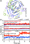
Fig. 2. The Cα RMSD (root-of-mean-squared-deviation) of RT from the coordinates in the reference X-ray structure in holoRT and in apoRT simulations. (a) A ribbon representation of the RT structure with the β-barrels colored in iceblue and the NT- and CT-barrels labelled. The catalytic triad and oxyanion hole at the β-barrel interface are shown in licorice. The Ca2+ ion is shown in a ball representation. (b) The Cα RMSD of catalytic triad and oxyanion hole residues (top), the β-barrel residues (middle), and the surface loops exhibiting active site occlusion upon BPTI unbinding and the Ca2+-binding loop (bottom).
Fig. 3
Fig. 3. Reorganization of the mechanical coupling network in RT upon BPTI unbinding. (a) The prominent mechanical responses of inter-residue couplings—top 25 percentile of the |ΔkIJ| values in the union list of the strongly coupled pairs in each rigidity graph, (square), (triangle), and (circle). The levels of softening (ΔkIJ < 0, blue) and stiffening (ΔkIJ > 0, red) responses are represented by the color bar. The prominent couplings with BPTI in holoRT are labelled on the diagonal. The key RT residue interacting with BPTI that starts a specific route of mechanical relay is used to annotate the chains of interaction network reorganization. If kIJ = 0 in the response state, the pair is labelled “off ” with ×. If kIJ = 0 in the reference state, the pair is labelled “on” with +. (b) The residues of mechanical relay systems in (a) on the RT structure.
Fig. 4
Fig. 4. Functional and evolutionary significance of protein intramolecular mechanical relays. Mechanically responsive residues in molecule-binding induced reorganization are mostly functional sites identified in experiments (red star), or have high sequence conservation (green star). The mechanical relay systems identified in Fig. 3 for RT and in Fig. 6 for PDZ3 are listed here on the primary sequence with secondary structures annotated. (a) In RT, H57, D102, S195, and G193 are the catalytic triad and oxyanion hole, respectively; nearby D189 and S214 are the S1 site for specificity control in substrate binding; R117 and S146 are protease autolysis sites; H40, E70, and W215 (ref. 43–45) at regulatory loop edges exhibit long-range effects on activities; R96 in a surface loop conducts active site occlusion in a homolog; with I16 and D194 activators forming a salt bridge, the activation domain involves N143 in l8, D189-C191 in l10, and C220 in l11; and functional mutation sensitivity has been demonstrated for the other labelled residues. (b) In PDZ3, the identified functional sites showing mutation sensitivity are mostly in the β-sandwish core. Including CT-extension in MSA as motivated by their inter-domain couplings in the rigidity graphs reveals the highly conserved residues shown here.
Fig. 5
Fig. 5. The Cα RMSD (root-of-mean-squared-deviation) of PDZ3 from the coordinates in the reference X-ray structure in holoPDZ3 and apoPDZ3 simulations. (a) A ribbon representation of the PDZ3 structure with the β-sandwich colored in iceblue. The CT extension is colored green and the CRIPT peptide is colored gray. (b) The Cα RMSD of β-sandwich (top), the α3-helix in CT-extension (middle), and β7–β8 hairpin in CT-extension (bottom).
Fig. 6
Fig. 6. Reorganization of the mechanical coupling network in PDZ3 upon binding the CRIPT peptide. (a) The prominent mechanical responses of inter-residue couplings—top 25 percentile of the |ΔkIJ| values in the union list of the strongly coupled pairs in each rigidity graph, (square), (triangle), and (circle). The levels of softening (ΔkIJ < 0, blue) and stiffening (ΔkIJ > 0, red) responses are represented by the color bar. The prominent couplings with CRIPT in holoPDZ3 are labelled on the diagonal. The key PDZ3 residue interacting with CRIPT that starts a specific route of mechanical relay is used to annotate the chains of interaction network reorganization. If kIJ = 0 in the response state, the pair is labelled off with ×. If kIJ = 0 in the reference state, the pair is labelled on with +. (b) The residues of mechanical relay systems in (a) on the PDZ3 structure. Definitions of sold/dash lines and arrows are as in Fig. 3(a). The S339-Y397 coupling prominent in both apoPDZ3 and holoPDZ3 is colored black.

Similar articles

Cited by

  • CHARMM at 45: Enhancements in Accessibility, Functionality, and Speed.
    Hwang W, Austin SL, Blondel A, Boittier ED, Boresch S, Buck M, Buckner J, Caflisch A, Chang HT, Cheng X, Choi YK, Chu JW, Crowley MF, Cui Q, Damjanovic A, Deng Y, Devereux M, Ding X, Feig MF, Gao J, Glowacki DR, Gonzales JE 2nd, Hamaneh MB, Harder ED, Hayes RL, Huang J, Huang Y, Hudson PS, Im W, Islam SM, Jiang W, Jones MR, Käser S, Kearns FL, Kern NR, Klauda JB, Lazaridis T, Lee J, Lemkul JA, Liu X, Luo Y, MacKerell AD Jr, Major DT, Meuwly M, Nam K, Nilsson L, Ovchinnikov V, Paci E, Park S, Pastor RW, Pittman AR, Post CB, Prasad S, Pu J, Qi Y, Rathinavelan T, Roe DR, Roux B, Rowley CN, Shen J, Simmonett AC, Sodt AJ, Töpfer K, Upadhyay M, van der Vaart A, Vazquez-Salazar LI, Venable RM, Warrensford LC, Woodcock HL, Wu Y, Brooks CL 3rd, Brooks BR, Karplus M. Hwang W, et al. J Phys Chem B. 2024 Oct 17;128(41):9976-10042. doi: 10.1021/acs.jpcb.4c04100. Epub 2024 Sep 20. J Phys Chem B. 2024. PMID: 39303207 Free PMC article. Review.
  • Statistical learning of protein elastic network from positional covariance matrix.
    Yu CC, Raj N, Chu JW. Yu CC, et al. Comput Struct Biotechnol J. 2023 Mar 28;21:2524-2535. doi: 10.1016/j.csbj.2023.03.033. eCollection 2023. Comput Struct Biotechnol J. 2023. PMID: 37095762 Free PMC article.
  • Mechanical codes of chemical-scale specificity in DNA motifs.
    Chen YT, Yang H, Chu JW. Chen YT, et al. Chem Sci. 2023 Aug 29;14(37):10155-10166. doi: 10.1039/d3sc01671d. eCollection 2023 Sep 27. Chem Sci. 2023. PMID: 37772098 Free PMC article.

References

    1. Changeux J. P. Science. 2005;308:1424–1428. doi: 10.1126/science.1108595. - DOI - PubMed
    1. Smock R. G. Gierasch L. M. Science. 2009;324:198–203. doi: 10.1126/science.1169377. - DOI - PMC - PubMed
    1. Latorraca N. R. Venkatakrishnan A. J. Dror R. O. Chem. Rev. 2016;117:139–155. doi: 10.1021/acs.chemrev.6b00177. - DOI - PubMed
    1. Cooper A. Dryden D. T. Eur. Biophys. J. 1984;11:103–109. doi: 10.1007/BF00276625. - DOI - PubMed
    1. Petit C. M. Zhang J. Sapienza P. J. Fuentes E. J. Lee A. L. Proc. Natl. Acad. Sci. U. S. A. 2009;106:18249–18254. doi: 10.1073/pnas.0904492106. - DOI - PMC - PubMed