

Comparison of endoscopic ultrasonography with and without contrast enhancement for characterization of pancreatic tumors: a meta-analysis
- PMID: 35433200
- PMCID: PMC9010094
- DOI: 10.1055/a-1782-5033
Comparison of endoscopic ultrasonography with and without contrast enhancement for characterization of pancreatic tumors: a meta-analysis
Abstract
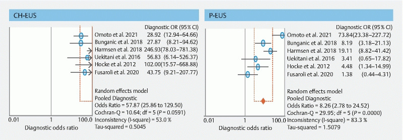
Background and study aims Endoscopic ultrasonography (EUS) is a reliable and efficient modality for detecting pancreatic tumors; however, plain EUS (P-EUS) is limited with respect to characterization of pancreatic tumors. Recently, the use of contrast-enhanced harmonic EUS (CH-EUS) has increased, and its utility for characterization of pancreatic tumors has been reported. This meta-analysis compares the diagnostic ability of P-EUS with that of CH-EUS for characterization of pancreatic tumors. Methods A systematic meta-analysis of all potentially relevant articles in PubMed, the Cochrane library, and Google Scholar databases was performed. Fixed effects or random effects models were used to investigate pooled sensitivity, specificity, positive likelihood ratio, and negative likelihood ratio, with 95 % confidence intervals (CIs). Results This meta-analysis included 719 patients who underwent CH-EUS and 723 who underwent P-EUS, from six eligible studies. The pooled estimates of sensitivity, specificity, and diagnostic odds ratio were 93 % (95 % CI, 0.90-0.95), 80 % (95 % CI, 0.75-0.85), and 57.9 (95 % CI, 25.9-130), respectively, for CH-EUS, and 86 % (95 % CI, 0.82-0.89), 59 % (95 % CI, 0.52-0.65), and 8.3 (95 % CI, 2.8-24.5) for P-EUS. The areas under the summary receiver operating characteristics curves for CH-EUS and P-EUS were 0.96 and 0.80, respectively. The diagnostic odds ratio for pancreatic cancer was 2.98 times higher on CH-EUS than on P-EUS ( P = 0.03). Funnel plots demonstrated no publication bias. Conclusions This meta-analysis demonstrates that CH-EUS has higher diagnostic accuracy for pancreatic cancer than P-EUS, and is thus a valuable tool for characterization of pancreatic tumors.
The Author(s). This is an open access article published by Thieme under the terms of the Creative Commons Attribution-NonDerivative-NonCommercial License, permitting copying and reproduction so long as the original work is given appropriate credit. Contents may not be used for commercial purposes, or adapted, remixed, transformed or built upon. (https://creativecommons.org/licenses/by-nc-nd/4.0/).
Conflict of interest statement
Competing interests Kitano and Napoléon have received honoraria from Olympus Corporation for presentations at conferences. Dr. Dietrich has received honoraria from Hitachi and Pentax Corporations for presentations at conferences.
Figures


References
-
- American Cancer Society . Atlanta: American Cancer Society; 2017. Cancer Facts & Figures 2017.
-
- The Editorial Board of the Cancer Statics in Japan, Foundation for Promotion of Cancer Research . Tokyo, Japan: FPCR c/o National Cancer Center; 2016. Cancer Statics in Japan-2015; pp. 1–129.
-
- Ahmad N A, Kochman M L, Lewis J D et al.Endosonography is superior to angiography in the preoperative assessment of vascular involvement among patients with pancreatic carcinoma. J Clin Gastroenterol. 2001;32:54–58. - PubMed
-
- DeWitt J, Devereaux B, Chriswell M et al.Comparison of endoscopic ultrasonography and multidetector computed tomography for detecting and staging pancreatic cancer. Ann Intern Med. 2004;141:753–763. - PubMed
Publication types
LinkOut - more resources
Full Text Sources