

The Cellular and Molecular Interaction Between Erythrocytes and Plasmodium falciparum Merozoites
- PMID: 35433504
- PMCID: PMC9008539
- DOI: 10.3389/fcimb.2022.816574
The Cellular and Molecular Interaction Between Erythrocytes and Plasmodium falciparum Merozoites
Abstract
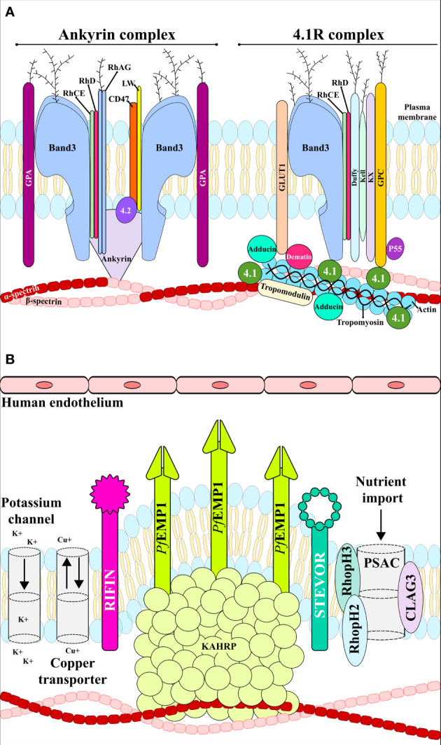
Plasmodium falciparum is the most lethal human malaria parasite, partly due to its genetic variability and ability to use multiple invasion routes via its binding to host cell surface receptors. The parasite extensively modifies infected red blood cell architecture to promote its survival which leads to increased cell membrane rigidity, adhesiveness and permeability. Merozoites are initially released from infected hepatocytes and efficiently enter red blood cells in a well-orchestrated process that involves specific interactions between parasite ligands and erythrocyte receptors; symptoms of the disease occur during the life-cycle's blood stage due to capillary blockage and massive erythrocyte lysis. Several studies have focused on elucidating molecular merozoite/erythrocyte interactions and host cell modifications; however, further in-depth analysis is required for understanding the parasite's biology and thus provide the fundamental tools for developing prophylactic or therapeutic alternatives to mitigate or eliminate Plasmodium falciparum-related malaria. This review focuses on the cellular and molecular events during Plasmodium falciparum merozoite invasion of red blood cells and the alterations that occur in an erythrocyte once it has become infected.
Keywords: host–parasite interaction; invasion; malaria; merozoite; pathogenesis; remodelling.
Copyright © 2022 Molina-Franky, Patarroyo, Kalkum and Patarroyo.
Conflict of interest statement
The authors declare that the research was conducted in the absence of any commercial or financial relationships that could be construed as a potential conflict of interest.
Figures


Similar articles
-
Deletion of the Plasmodium falciparum merozoite surface protein 7 gene impairs parasite invasion of erythrocytes.Eukaryot Cell. 2008 Dec;7(12):2123-32. doi: 10.1128/EC.00274-08. Epub 2008 Sep 26. Eukaryot Cell. 2008. PMID: 18820076 Free PMC article.
-
Binding of aldolase and glyceraldehyde-3-phosphate dehydrogenase to the cytoplasmic tails of Plasmodium falciparum merozoite duffy binding-like and reticulocyte homology ligands.mBio. 2012 Sep 18;3(5):e00292-12. doi: 10.1128/mBio.00292-12. Print 2012. mBio. 2012. PMID: 22991428 Free PMC article.
-
Revealing the sequence and resulting cellular morphology of receptor-ligand interactions during Plasmodium falciparum invasion of erythrocytes.PLoS Pathog. 2015 Feb 27;11(2):e1004670. doi: 10.1371/journal.ppat.1004670. eCollection 2015 Feb. PLoS Pathog. 2015. PMID: 25723550 Free PMC article.
-
RBC membrane biomechanics and Plasmodium falciparum invasion: probing beyond ligand-receptor interactions.Trends Parasitol. 2022 Apr;38(4):302-315. doi: 10.1016/j.pt.2021.12.005. Epub 2022 Jan 4. Trends Parasitol. 2022. PMID: 34991983 Free PMC article. Review.
-
Erythrocyte glycophorins as receptors for Plasmodium merozoites.Parasit Vectors. 2019 Jun 24;12(1):317. doi: 10.1186/s13071-019-3575-8. Parasit Vectors. 2019. PMID: 31234897 Free PMC article. Review.
Cited by
-
Plasmodium Falciparum and mosquito vector IgG patterns across suspected malaria cases in Ghana.BMC Infect Dis. 2024 Dec 2;24(1):1374. doi: 10.1186/s12879-024-10248-9. BMC Infect Dis. 2024. PMID: 39623362 Free PMC article.
-
Healthcare-associated malaria: a systematic review, 1997 to 2023.Euro Surveill. 2025 Mar;30(11):2400393. doi: 10.2807/1560-7917.ES.2025.30.11.2400393. Euro Surveill. 2025. PMID: 40116034 Free PMC article.
-
Red Blood Cells Oligosaccharides as Targets for Plasmodium Invasion.Biomolecules. 2022 Nov 11;12(11):1669. doi: 10.3390/biom12111669. Biomolecules. 2022. PMID: 36421683 Free PMC article. Review.
-
PLASMOpred: A Machine Learning-Based Web Application for Predicting Antimalarial Small Molecules Targeting the Apical Membrane Antigen 1-Rhoptry Neck Protein 2 Invasion Complex.Pharmaceuticals (Basel). 2025 May 23;18(6):776. doi: 10.3390/ph18060776. Pharmaceuticals (Basel). 2025. PMID: 40573173 Free PMC article.
-
Ontological representation, modeling, and analysis of parasite vaccines.J Biomed Semantics. 2024 Apr 25;15(1):4. doi: 10.1186/s13326-024-00307-0. J Biomed Semantics. 2024. PMID: 38664818 Free PMC article.
References
Publication types
MeSH terms
Substances
LinkOut - more resources
Full Text Sources
Medical