. 2022 Mar 30:12:826192.
doi: 10.3389/fcimb.2022.826192. eCollection 2022.

Immunomodulation as a Protective Strategy in Chronic Otitis Media

Affiliations

Immunomodulation as a Protective Strategy in Chronic Otitis Media

Anke Leichtle et al. Front Cell Infect Microbiol. .

Abstract

Introduction: Major features of the pathogenesis in otitis media, the most common disease in childhood, include hyperplasia of the middle ear mucosa and infiltration by leukocytes, both of which typically resolve upon bacterial clearance via apoptosis. Activation of innate immune receptors during the inflammatory process leads to the activation of intracellular transcription factors (such as NF-κB, AP-1), which regulate both the inflammatory response and tissue growth. We investigated these leading signaling pathways in otitis media using mouse models, human samples, and human middle ear epithelial cell (HMEEC) lines for therapeutic immunomodulation.

Methods: A stable otitis media model in wild-type mice and immunodeficient KO-mice, as well as human tissue samples from chronic otitis media, skin from the external auditory canal and middle ear mucosa removed from patients undergoing ear surgery, were studied. Gene and protein expression of innate immune signaling molecules were evaluated using microarray, qPCR and IHC. In situ apoptosis detection determined the apoptotic rate. The influence of bacterial infection on immunomodulating molecules (TNFα, MDP, Tri-DAP, SB203580, Cycloheximide) in HMEEC was evaluated. HMEEC cells were examined after bacterial stimulation/inhibition for gene expression and cellular growth.

Results: Persistent mucosal hyperplasia of the middle ear mucosa in chronic otitis media resulted from gene and protein expression of inflammatory and apoptotic genes, including NODs, TNFα, Casp3 and cleaved Casp3. In clinical chronic middle ear samples, these molecules were modulated after a specific stimulation. They also induced a hyposensitive response after bacterial/NOD-/TLR-pathway double stimulation of HMEEC cells in vitro. Hence, they might be suitable targets for immunological therapeutic approaches.

Conclusion: Uncontrolled middle ear mucosal hyperplasia is triggered by TLRs/NLRs immunoreceptor activation of downstream inflammatory and apoptotic molecules.

Keywords: NOD-like receptors; TNFα; TOLL-like receptors; apoptosis; immunology; otitis media.


Conflict of interest statement

AR is a co-founder of Otonomy Inc., serves as a member of the Scientific Advisory Board, and holds an equity position in the company. The UCSD Committee on Conflict of Interest has approved this relationship. Otonomy, Inc. played no part in the research reported here. The remaining authors declare that the research was conducted in the absence of any commercial or financial relationships that could be construed as a potential conflict of interest.

Figures

Figure 1
Figure 1
Innate immune signaling pathways implicated in bacterial Otitis Media (OM). The most common receptors involved in pattern recognition are the Toll-like receptors (TLRs), and NOD-like receptors (NRLs) of which signaling leads to the activation of Nuclear factor of kappa-B (NF-κB), the main transcription factor involved in cytokine production. Stimulation of NLRP3 leads the inflammasome to recruit and activate caspase-1, which in turn cleaves the pro-forms of IL1ß, Il18. These pro-inflammatory cytokines are in turn released in their mature/bioactive forms. The intracellular Nucleotide oligomerization domain -1 and -2 (NODs 1 and 2) can also induce inflammation through the RICK/RIP2 pathway. However, NOD2 can also induce autophagy through ATG16L1 shifting the balance of NOD2 downstream signaling. Crosstalk between the different immune elements: TLRs/NRLs, the Inflammasome and Growth factors are believed to modulate the immune responses.
Figure 2
Figure 2
Assessment of genes involved in patter recognition during a complete episode of acute NTHi-induced OM in the mouse, from initiation to recovery, evaluated by gene arrays. The cell surface TLR2, TLR4 and TLR6 were expressed in sentinel mucosa and highly upregulated in early time points (3hrs to 24hrs). The intracellular PRRs TLR9 was upregulated at later time points (2d). Meanwhile, NLRP3 expression peaked between (3hrs to 24hrs). The DNA-dependent activator of IFN-regulatory factors (DAI) and DNA sensor RNA polymerase (Pol-III) expression peaked later at day 2 onwards. *p < .05; **P < .01
Figure 3
Figure 3
Pathophysiology of the middle ear during a NTHi induced OM in WT mice (C57/BL6). (A) OM time course showing cross-sections of the ME bulla and revealing that the ME mucosal hyperplasia and cellular infiltration peaks at 2-3 days after bacterial inoculation and recover by 5-7 days. Cellular infiltrate are evident on day 1, peak by day 2 and return to baseline by day 7 (B) Middle ear mucosa thickening is seen at a higher magnification (10x) at the indicated OM time points.
Figure 4
Figure 4
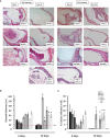
Representative histological changes at 3 and 10 days post NTHi challenge seen in middle ears of different immunodeficient KO mice. (A) Considerable inflammation in the ME space (filling the cavity with fluid and inflammatory cells) is observed on day 3 and by day 10 persistent mucosal hyperplasia and cell recruitment is noted in some of the immunodeficient KO animals. (B) Quantitative analysis of mucosal thickness and (C) the % area of the middle ear cavity covered with inflammatory cells in KO mice. (n=6 ears, *p < 0.05).
Figure 5
Figure 5
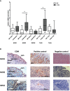
(A) Gene expression of Innate Immune Receptors in human COM. mRNA expression of NOD1, NOD2, RIPK2, TLR2 and TLR4 in healthy middle ear samples (ME) and COM relative to GAPDH. Significantly increased gene expression of NOD2 and TLR2 in COM compared to healthy middle ear tissue. For normalization, the housekeeping gene GAPDH. N = 20 each; GraphPad Prism with an unpaired t-test, *p < .05. (B) Protein expression of NLR Immune Receptors in human COM. NOD2, NOD1 and RIPK2 could be localized immunohistochemically mainly intra- and subepithelial in COM. The right column displays their positive control in small intestine (NOD1) and in pancreas (NOD2 and RIPK2). Orange/brown represents the target molecule NOD1, NOD2 and RIPK2, blue represents nuclei. Scale is shown by scale bar insert.
Figure 6
Figure 6
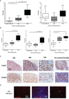
(A) Gene expression of TNF and IL8 in human COM. TNFα (left) and IL8 (right) mRNA expression in healthy middle ear samples, external auditory skin (EAS) and COM relative to GAPDH. Significantly increased gene expression of TNFα in COM compared to EAS and healthy middle ear samples. No significant mRNA regulation of IL8. (B) Gene expression of apoptotic genes in human COM. Bid, Casp3, and Casp7 mRNA expression in healthy middle ear samples, external auditory skin (EAS) and COM relative to GAPDH. Bid and Casp3 mRNA expression in COM is significantly increased to EAS and healthy middle ear samples (left and middle). Significant upregulation of Casp7 in EAS and COM to healthy samples (right). Normalization with GAPDH; N = 20 each; GraphPad Prism with an unpaired t-test, *p < .05. (C) Expression of apoptotic proteins. Immunohistochemistry by immunoperoxidase of Casp3 and cCasp3 as brownish-orange staining in human COM. Casp3 is broadly stained in the epithelium and subepithelial, whereas active cCasp3 protein expression shows staining mainly in the subepithelial layers. 20x, 40x and 63x magnification. (D) Caspase3 and 7 livestain in COM with cell effusion shows a disseminated red staining of single cells mainly subepithelial. 20x and 63x magnification.
Figure 7
Figure 7
Immunmodulation in the Human Middle Ear Epithelial Cell Line (HMEEC). Gene expression of TNF (A) and IL1β (B) in HMECC at 6h and 24 h after stimulation with NTHi, TNF, Tri-DAP, MDP, SB203580, CHX or double-stimulation, was evaluated using qPCR. For normalization, the housekeeping gene GAPDH was used. GraphPad Prism with an unpaired t-test, *p<0.05. (A) Increased gene expression of TNFα after stimulation with NTHi, TNFα and CHX or their double stimulation with NTHi at 6h in HMEEC compared to medium. At 24h induction of all stimuli or their double stimulation with NTHi in HMEEC compared to medium. However, double-stimulation with NTHi and TNFα, or Tri-DAP, or MDP, or SB203580 compared to the direct single stimuli molecule showed hyporesponse at 24h, with a reduction in TNFα expression (red arrows). (B) Significantly increased gene expression of IL1β only after stimulation with single stimulation with NTHi, or CHX; or double stimulation with NTHi and TNFα, TriDAP, MDP and CHX at 6h compared to controls. At 24h significant IL1β induction was observed under all conditions compared to controls. However, double-stimulation with NTHi and TNFα, Tri-DAP, MDP or SB203580 responded with no change in IL1β gene expression when compared to stimulation with NTHi alone at 24h. (C) Effects of NTHi, TNF, TriDAP, MDP and their combinations on the cellular viability of cultured HMEECs assessed by MTT assay. TNFα had a significant effect on cellular viability after incubation with NTHi Significant difference (p < 0.05) is marked with *.

References

    1. Aggarwal B. B. (2003). Signalling Pathways of the TNF Superfamily: A Double-Edged Sword. Nat. Rev. Immunol. 3 (9), 745–756. doi: 10.1038/nri1184 - DOI - PubMed
    1. Baltimore D., Beg A. A. (1995). DNA-Binding Proteins. A Butterfly Flutters by. Nature 373 (6512), 287–288. doi: 10.1038/373287a0 - DOI - PubMed
    1. Birkle T., Brown G. C. (2021). I’m Infected, Eat Me! Innate Immunity Mediated by Live, Infected Cells Signaling To Be Phagocytosed. Infection Immun. 89 (5), e00476–20. doi: 10.1128/IAI.00476-20 - DOI - PMC - PubMed
    1. Chen G., Goeddel D. V. (2002). TNF-R1 Signaling: A Beautiful Pathway. Science 296 (5573), 1634–1635. doi: 10.1126/science.1071924 - DOI - PubMed
    1. Chonmaitree T., Alvarez-Fernandez P., Jennings K., Trujillo R., Marom T., Loeffelholz M. J., et al. . (2015). Symptomatic and Asymptomatic Respiratory Viral Infections in the First Year of Life: Association With Acute Otitis Media Development. Clin. Infect. Dis. 60 (1), 1–9. doi: 10.1093/cid/ciu714 - DOI - PMC - PubMed

Publication types