Computed tomography perfusion imaging evaluation of angiogenesis in patients with pancreatic adenocarcinoma
- PMID: 35434057
- PMCID: PMC8968604
- DOI: 10.12998/wjcc.v10.i8.2393
Computed tomography perfusion imaging evaluation of angiogenesis in patients with pancreatic adenocarcinoma
Abstract
Background: Pancreatic adenocarcinoma is one of the most common malignant tumors of the digestive system. More than 80% of patients with pancreatic adenocarcinoma are not diagnosed until late stage and have distant or local metastases.
Aim: To investigate the value of computed tomography (CT) perfusion imaging in the evaluation of angiogenesis in pancreatic adenocarcinoma patients.
Methods: This is a retrospective cohort study. Patients with pancreatic adenocarcinoma and volunteers without pancreatic diseases underwent CT perfusion imaging from December 2014 to August 2017 in Huashan Hospital, Fudan University Shanghai, China.
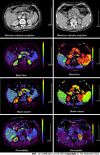
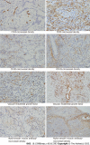
Results: A total number of 35 pancreatic adenocarcinoma patients and 33 volunteers were enrolled. The relative blood flow (rBF), and relative blood volume (rBV) were significantly lower in patients with pancreatic adenocarcinoma than in the control group (P < 0.05). Conversely, the relative permeability in patients with pancreatic adenocarcinoma was significantly higher than that in controls (P < 0.05). In addition, rBF, rBV, and the vascular maturity index (VMI) were significantly lower in grade III-IV pancreatic adenocarcinoma than in grade I-II pancreatic adenocarcinoma (P < 0.05). Vascular endothelial growth factor (VEGF), CD105-MVD, CD34-MVD, and angiogenesis rate (AR) were significantly higher in grade III-IV pancreatic adenocarcinoma than in grade I-II pancreatic adenocarcinoma (P < 0.05). Significant correlations between rBF and VEGF, CD105-MVD, AR, and VMI (P < 0.01) were observed. Moreover, the levels of rBV were statistically significantly correlated with those of VEGF, CD105-MVD, CD34-MVD, and VMI (P < 0.01).
Conclusion: Perfusion CT imaging may be an appropriate approach for quantitative assessment of tumor angiogenesis in pancreatic adenocarcinoma.
Keywords: Angiogenesis; Evaluation; Imaging; Pancreatic adenocarcinoma; Perfusion computed tomography; Quantitative assessment.
©The Author(s) 2022. Published by Baishideng Publishing Group Inc. All rights reserved.
Conflict of interest statement
Conflict-of-interest statement: The authors have declared that they have no competing interests.
Figures


Similar articles
-
Peripheral pulmonary nodules: relationship between multi-slice spiral CT perfusion imaging and tumor angiogenesis and VEGF expression.BMC Cancer. 2008 Jun 30;8:186. doi: 10.1186/1471-2407-8-186. BMC Cancer. 2008. PMID: 18590539 Free PMC article.
-
[Biological relativity of imaging of CT perfusion for pancreases to pancreatic cancer].Sichuan Da Xue Xue Bao Yi Xue Ban. 2009 May;40(3):521-4. Sichuan Da Xue Xue Bao Yi Xue Ban. 2009. PMID: 19627019 Chinese.
-
Quantification of angiogenesis by CT perfusion imaging in liver tumor of rabbit.Hepatobiliary Pancreat Dis Int. 2009 Apr;8(2):168-73. Hepatobiliary Pancreat Dis Int. 2009. PMID: 19357031
-
Evaluation of angiogenesis in colorectal carcinoma with multidetector-row CT multislice perfusion imaging.Eur J Radiol. 2010 Aug;75(2):191-6. doi: 10.1016/j.ejrad.2009.04.058. Epub 2009 May 29. Eur J Radiol. 2010. PMID: 19481397
-
Colorectal tumor vascularity: quantitative assessment with multidetector CT--do tumor perfusion measurements reflect angiogenesis?Radiology. 2008 Nov;249(2):510-7. doi: 10.1148/radiol.2492071365. Epub 2008 Sep 23. Radiology. 2008. PMID: 18812560
Cited by
-
Can quantitative perfusion CT-based biomarkers predict renal cell carcinoma subtypes?Abdom Radiol (NY). 2025 Jun;50(6):2586-2594. doi: 10.1007/s00261-024-04746-2. Epub 2024 Dec 17. Abdom Radiol (NY). 2025. PMID: 39688674
References
-
- Hezel AF, Kimmelman AC, Stanger BZ, Bardeesy N, Depinho RA. Genetics and biology of pancreatic ductal adenocarcinoma. Genes Dev. 2006;20:1218–1249. - PubMed
-
- Kim SI, Shin JY, Park JS, Jeong S, Jeon YS, Choi MH, Choi HJ, Moon JH, Hwang JC, Yang MJ, Yoo BM, Kim JH, Lee HW, Kwon CI, Lee DH. Vascular enhancement pattern of mass in computed tomography may predict chemo-responsiveness in advanced pancreatic cancer. Pancreatology. 2017;17:103–108. - PubMed
LinkOut - more resources
Full Text Sources
Research Materials

