. 2022 Jul;9(3):032211.
doi: 10.1117/1.NPh.9.3.032211. Epub 2022 Apr 7.

Toward an integrative neurovascular framework for studying brain networks

Affiliations

Toward an integrative neurovascular framework for studying brain networks

Jérémie Guilbert et al. Neurophotonics. 2022 Jul.

Abstract

Brain functional connectivity based on the measure of blood oxygen level-dependent (BOLD) functional magnetic resonance imaging (fMRI) signals has become one of the most widely used measurements in human neuroimaging. However, the nature of the functional networks revealed by BOLD fMRI can be ambiguous, as highlighted by a recent series of experiments that have suggested that typical resting-state networks can be replicated from purely vascular or physiologically driven BOLD signals. After going through a brief review of the key concepts of brain network analysis, we explore how the vascular and neuronal systems interact to give rise to the brain functional networks measured with BOLD fMRI. This leads us to emphasize a view of the vascular network not only as a confounding element in fMRI but also as a functionally relevant system that is entangled with the neuronal network. To study the vascular and neuronal underpinnings of BOLD functional connectivity, we consider a combination of methodological avenues based on multiscale and multimodal optical imaging in mice, used in combination with computational models that allow the integration of vascular information to explain functional connectivity.

Keywords: BOLD fMRI; brain optical imaging; functional connectivity; neurovascular coupling; neurovascular networks; vasculo-neuronal interactions.


Figures

Fig. 1
Fig. 1
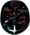
Vascular influences on BOLD FC. Functional connections observed through BOLD fMRI can be influenced in various ways by the vasculature. Nodes and edges depict elements of a BOLD functional network. Propagation delays of blood-borne signals can induce time lags which reduce correlations or generate spurious ones. Physiological oscillations can create non-neuronal correlations which may be accentuated in strongly vascularized regions such as the occipital cortex, making it harder to detect neuronal correlations. CBF is increased in hub regions where metabolic requirements are heightened, highlighting a local form of coupling between vascular and neuronal organization. Furthermore, vascular properties are thought to be organized in spatially remote areas to functionally match RSNs. The resulting coordinated delivery of blood in RSNs leads to observations in fMRI of spatial components associated with purely vascular signals in addition to neuronally-driven ones.
Fig. 2
Fig. 2
Imaging neurovascular networks at different spatial scales. The interactions between neurons and vasculature can be observed in vivo at different spatial scales. At the microscopic scale, calcium imaging in neurons at cellular resolution can be combined with colocalized imaging of blood vessels and other cell types within the NVU. At the mesoscopic scale, widefield calcium imaging can be combined with vascular optical measurements in cortical surface vessels. At the macroscopic scale, BOLD fMRI measures entangled neuronal and vascular interactions. Structural connections are measured using diffusion MRI and regional blood flow using arterial spin labeling. Small pictograms depict the translational perspective of macroscopic noninvasive brain imaging, whereas smaller scales are only accessible in animal models. Bridging across scales can be accomplished experimentally with multiscale measurements in a single animal. Compiling results from standardized measurement protocols repeated across entire brains can yield statistically representative maps of microscopic properties, or atlases, to which macroscopic datasets can be coregistered.
Fig. 3
Fig. 3
Divergence between BOLD FC and neuronal FC. Although a good agreement is generally observed between FC derived from BOLD and from calcium measurements, the cell-type specificity of neurovascular coupling can potentially lead to divergence between the two, as observed in Ref. . Specifically in the mouse barrel cortex, the expected positive relationship between connectivity strength evaluated with BOLD and excitatory calcium signals holds only when considering interhemispheric nodes (left upper panel). When considering intrahemispheric nodes, the relationship is negative (left lower panel). Taking into account the likely presence of strong interhemispheric inhibition in the barrel cortex, this discrepancy can be explained by positing that inhibitory populations can simultaneously generate positive BOLD signals while inhibiting excitatory neurons across hemispheres. The arrows in the right panel represent the relationships between the different networks, with hemoneural interactions possibly mediating a link between the vascular and functional neuronal networks. The left panel is a sketch of the results from Ref. .

References

    1. Ogawa S., et al. , “Brain magnetic resonance imaging with contrast dependent on blood oxygenation,” Proc. Natl. Acad. Sci. U. S. A. 87, 9868–9872 (1990).10.1073/pnas.87.24.9868 - DOI - PMC - PubMed
    1. Iadecola C., “The neurovascular unit coming of age: a journey through neurovascular coupling in health and disease,” Neuron 96, 17–42 (2017).NERNET10.1016/j.neuron.2017.07.030 - DOI - PMC - PubMed
    1. Hillman E. M. C., “Coupling mechanism and significance of the BOLD signal: a status report,” Annu. Rev. Neurosci. 37, 161–181 (2014).ARNSD510.1146/annurev-neuro-071013-014111 - DOI - PMC - PubMed
    1. Tong Y., et al. , “Can apparent resting state connectivity arise from systemic fluctuations?” Front. Hum. Neurosci. 9, 285 (2015).10.3389/fnhum.2015.00285 - DOI - PMC - PubMed
    1. Bright M. G., et al. , “Vascular physiology drives functional brain networks,” NeuroImage 217, 116907 (2020).NEIMEF10.1016/j.neuroimage.2020.116907 - DOI - PMC - PubMed