. 2022 Mar 25;25(4):104153.
doi: 10.1016/j.isci.2022.104153. eCollection 2022 Apr 15.

A dual SHOX2:GFP; MYH6:mCherry knockin hESC reporter line for derivation of human SAN-like cells

Affiliations

A dual SHOX2:GFP; MYH6:mCherry knockin hESC reporter line for derivation of human SAN-like cells

Zaniar Ghazizadeh et al. iScience. .

Abstract

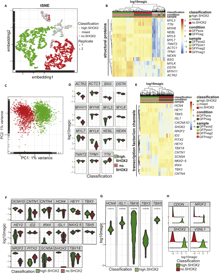
The sinoatrial node (SAN) is the primary pacemaker of the heart. The human SAN is poorly understood due to limited primary tissue access and limitations in robust in vitro derivation methods. We developed a dual SHOX2:GFP; MYH6:mCherry knockin human embryonic stem cell (hESC) reporter line, which allows the identification and purification of SAN-like cells. Using this line, we performed several rounds of chemical screens and developed an efficient strategy to generate and purify hESC-derived SAN-like cells (hESC-SAN). The derived hESC-SAN cells display molecular and electrophysiological characteristics of bona fide nodal cells, which allowed exploration of their transcriptional profile at single-cell level. In sum, our dual reporter system facilitated an effective strategy for deriving human SAN-like cells, which can potentially be used for future disease modeling and drug discovery.

Keywords: Biological sciences; Methodology in biological sciences; Stem cells research.


Conflict of interest statement

T.E. and S.C. are founding owners of OncoBeat, LLC. A patent has been filled for “Compositions and methods for generation of sinoatrial node-like cells and their use in drug discovery”.

Figures

None
Graphical abstract
Figure 1
Figure 1
Generation of human SAN-like cells from hESCs (A) Schematic representation of the differentiation protocols 1–8. The protocol involves three stages (S1, S2, and S3) during the first 9 days of directed differentiation. (B–D) Quantitative PCR (B), live fluorescence images (C), and FACS plots (D) of the day 30 cells derived from the SHOX2:GFP;MYH6:mCherry H9 line using the corresponding differentiation protocols (n = at least 3 biological replicates for each condition). For quantitative PCR data, fold changes were normalized to protocol #1. p values calculated using ordinary one-way ANOVA with Fisher’s test (∗∗p < 0.01, ∗∗∗p < 0.001). Data are represented as mean ± SEM. The x axis in panel D is side scatter. (E) Quantitative PCR analysis for SHOX2 transcriptional expression of GFP+ cells purified after sorting. n = 3 biological replicates. p value calculated by unpaired two-tailed Student’s t-test ∗∗∗p < 0.001. Data are represented as mean ± SEM, and normalized to GFP-negative population. (F) Schematic and results of the epigenetic library chemical screen. (G) Schematic of the final differentiation protocol. (H) Immunofluorescence image and FACS plot of day 30 cells derived using the protocol described in (G), Scale bar = 100 μm. AA: Activin A, B: BMP4, Ch: CHIR99021, Wi: Wnt inhibitor, RA: Retinoic Acid, Fi: FGF inhibitor, Cu: cucurbitacin, Tyr490: tyrphostin AG 490. See also Figures S1 and S2.
Figure 2
Figure 2
Transcriptome profiling of hESC-SAN cells (A and B) Hierarchical clustering (A) and heatmap (B) of transcriptional profiling in GFP+ cells and mCherry+ cells purified as described in (S2B) compared to murine SAN or murine right atrial (RA) profiles (GSE65658). (C) Gene set enrichment analysis in purified GFP+ cells and GFP-mCherry+ cells. Murine sinoatrial and atrial tissues (GSE65658) were used as controls. RA: murine right atrial sample; SAN: murine sinoatrial node sample. R1, R2, and R3 represent independent replicates and the labels P4 and P14 refer to post-natal day 4 and day 14 samples. The human SAN-like cell profiles always cluster with murine SAN profiles, while human CM cell profiles always cluster with murine right atrium myocardium profiles.
Figure 3
Figure 3
Functional characterization of hESC-SAN cells (A) Immunofluorescent co-staining of GFP with pacemaker-associated transcription factors (ISL1, TBX5, and NKX2.5) and ion channels (HCN4, Cav3.1, Cav1.3, and Cx30.2) in cells differentiated to day 30 or day 60. Scale bar = 200 μm. (B) qRT-PCR analysis using samples from GFP+ (G+) or mCherry+ (Ch+) cells at day 25 or day 45. n = 3 biological replicates. Fold change was normalized to undifferentiated hESCs. p values calculated by one-way ANOVA with Fisher’s test (∗p < 0.05, ∗∗p < 0.01, ∗∗∗p < 0.001). Data are represented as mean ± SEM. (C) Schematic for co-culture of hESC-derived ANs expressing eYFP-channelrhodopsin-2 with either hESC-SAN or hESC-V. (D) Representative immunofluorescence (left panel) and electron microscopy images (right panel) of hESC-AN and hESC-SAN after 12 weeks of co-culture. Scale bar: 100 μm (left panel) and 1 μm (right panel). (E) Basal release of catecholamine was analyzed using an ELISA kit for AN alone, AN + hESC-SAN and AN + hESC-V conditions after 8 weeks of co-culture. n = 3 biological replicates. p values calculated by one-way ANOVA (∗∗p < 0.01). Data are represented as mean ± SEM. (F) Representative image of averaged action potentials recorded by perforated patch clamp from individual hESC-SAN (left) and hESC-V (right) cells and distribution of dV/dtmax, APD30, and APD90 recorded used day 30 cells. p values calculated by one-way ANOVA used to compare means (∗p < 0.05). Data are represented as mean ± SEM. (G) Beating rate of hESC-SAN and hESC-V cells. p values calculated by unpaired two-tailed Student’s t-test ∗∗∗∗p < 0.0001. Data are represented as mean ± SEM. (H) Spontaneous firing of hESC-SAN cells. Note that for these experiments, cells were replated as single cells on cover slips and they were maintained in physiological buffers (rather than their optimal cell culture media). Cells are manipulated for up to an hour at room temperature, during which time the beating rate decreases substantially (compared to conditions in G). (I) Ih current measurements in hESC-SAN or hESC-V cells by patch clamp. Left: Example traces of hESC-SAN cells; Middle: Averaged I-V curves; Right: Example traces of hESC-V cells. (n = 5 biological replicates). (J) Quantitative analysis of day 30 hESC-SAN function in response to 1 μM ivabradine. Fold change in calcium transient amplitude (deltaF/F0) of hESC-SAN or hESC-V cells before and after treatment with ivabradine (n = 22 biological replicates for hESC-SAN and n = 17 biological replicates for hESC-V). Unpaired two-tailed Student’s t-test was used to compare means (∗p < 0.05). Each dot represents data measured from independent cells. (K) Quantitative analysis of day 30 hESC-SAN function in response to 1 μM isoproterenol and 5 μM carbachol. p values calculated by unpaired two-tailed Student’s t-test ∗p < 0.05, ∗∗p < 0.01. Data are represented as mean ± SEM. See also Figure S3A and Video S1.
Figure 4
Figure 4
Single-cell RNA sequencing analysis of differentiated hESC-SAN and hESC-V cells (A) t-SNE plots colored according to the cell populations. (B) Heatmap based on structural protein expression showing the clustering of the high-SHOX2, mixed-, and no- SHOX2 cells. (C) PCA plot of high-SHOX2 and no-SHOX2 cells. (D) Jitter plots based on structural protein expression of high-SHOX2 and no-SHOX2 cells. (E and F) Heatmap (E) and jitter plots (F) based on expression of key transcription factors and ion channels in high-SHOX2 and no-SHOX2 cells. (G and H) Jitter plots for the expression of HCN4, ISL1, TBX3, TBX5, and TBX18 (G) and jitter plots for the expression of SHOX2, CDON, NR2F2, and VSNL1, (H) in the high-SHOX2 and no-SHOX2 cells. See also Figure S3B.

References

    1. Birket M.J., Ribeiro M.C., Verkerk A.O., Ward D., Leitoguinho A.R., den Hartogh S.C., Orlova V.V., Devalla H.D., Schwach V., Bellin M., et al. Expansion and patterning of cardiovascular progenitors derived from human pluripotent stem cells. Nat. Biotechnol. 2015;33:970–979. - PubMed
    1. Burkhard S.B., Bakkers J. Spatially resolved RNA-sequencing of the embryonic heart identifies a role for Wnt/β-catenin signaling in autonomic control of heart rate. Elife. 2018;7 - PMC - PubMed
    1. Butler A., Hoffman P., Smibert P., Papalexi E., Satija R. Integrating single-cell transcriptomic data across different conditions, technologies, and species. Nat. Biotechnol. 2018;36:411–420. doi: 10.1038/nbt.4096. - DOI - PMC - PubMed
    1. Cingolani E., Goldhaber J.I., Marban E. Next-generation pacemakers: from small devices to biological pacemakers. Nat. Rev. Cardiol. 2017;15:139–150. - PMC - PubMed
    1. Cong L., Ran F.A., Cox D., Lin S., Barretto R., Habib N., Hsu P.D., Wu X., Jiang W., Marraffini L.A., et al. Multiplex genome engineering using CRISPR/Cas systems. Science. 2013;339:819–823. - PMC - PubMed