

Surgical Management of the Trapezium Canal Syndrome: An Uncommon Presentation of Tenosynovitis of Flexor Carpi Radialis
- PMID: 35434570
- PMCID: PMC9005376
- DOI: 10.1016/j.jhsg.2022.01.001
Surgical Management of the Trapezium Canal Syndrome: An Uncommon Presentation of Tenosynovitis of Flexor Carpi Radialis
Abstract
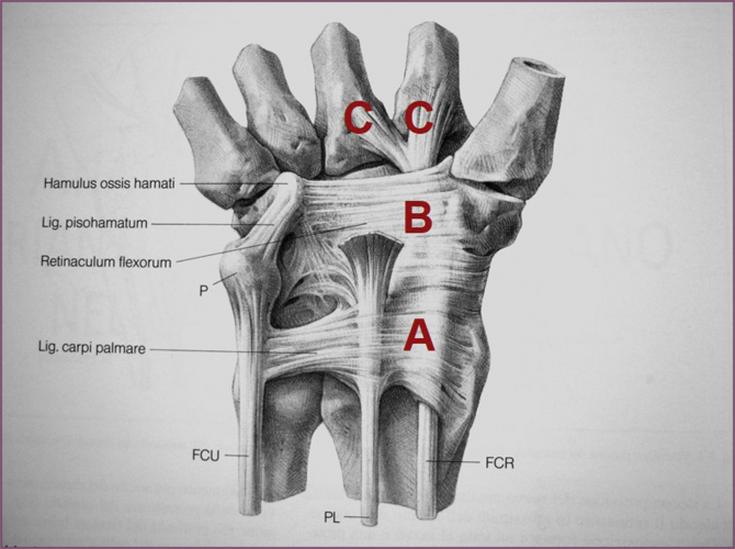
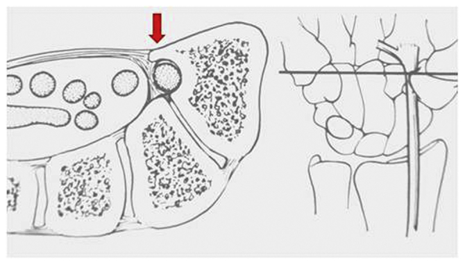
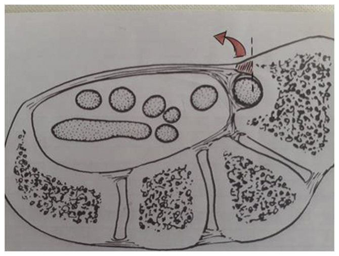
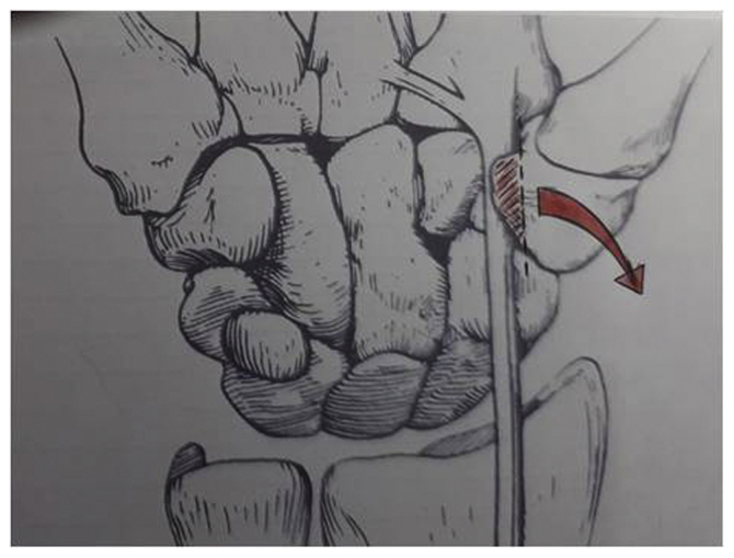
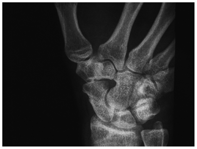
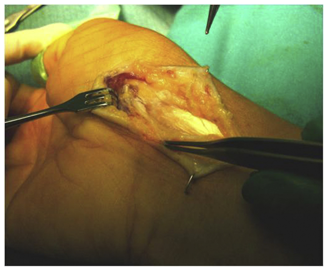
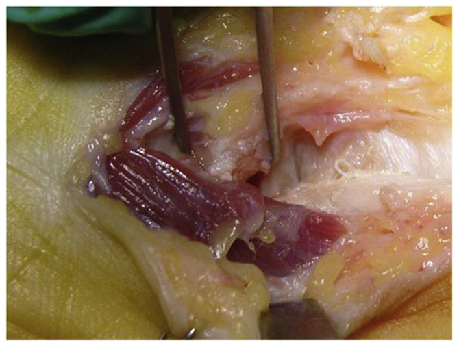
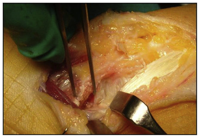
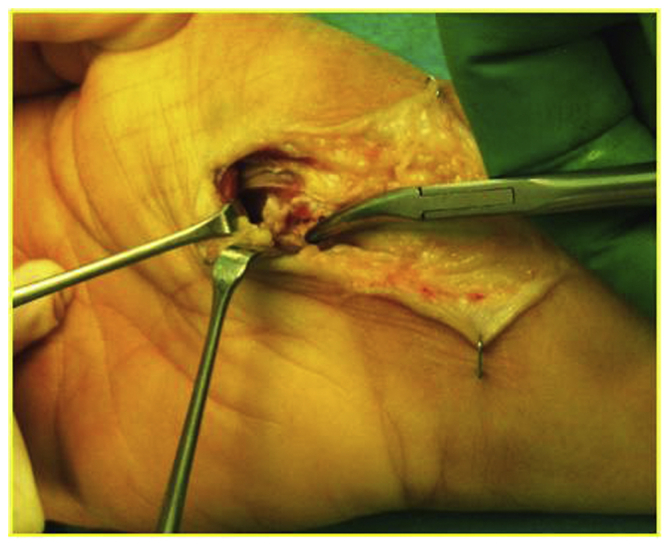
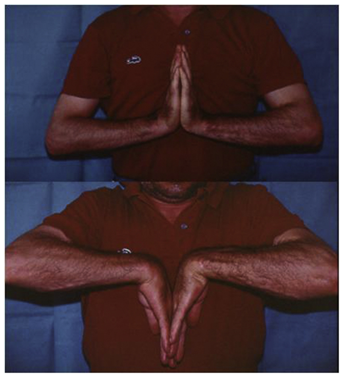
Flexor carpi radialis (FCR) tenosynovitis is a condition characterized by pain over the volar radial wrist caused by inflammation of the FCR tendon sheath. It is an uncommon and often unrecognized pathology that could be misleading from a diagnostic and therapeutic point of view. Treatment usually involves immobilization, nonsteroidal anti-inflammatory drugs, and injections. In refractory cases, operative release of the FCR tendon sheath may be indicated. In this article, we report our experience in treating FCR tenosynovitis by surgically decompressing the trapezium canal, through which the tendon runs, at the wrist. In our experience, this surgical technique allows a good functional recovery with the resolution of painful symptoms without notable complications.
Keywords: Flexor carpi radialis; Tenosynovitis; Trapezium canal.
© 2022 The Authors.
Figures
References
-
- Bishop A.T., Gabel G., Carmichael S.W. Flexor carpi radialis tendinitis. Part I: operative anatomy. J Bone Joint Surg Am. 1994;76(7):1009–1014. - PubMed
-
- Nigro R.O. Anatomy of the flexor retinaculum of the wrist and the flexor carpi radialis tunnel. Hand Clin. 2001;17(1):61–64. vi. - PubMed
-
- Adams J.E., Habbu R. Tendinopathies of the hand and wrist. J Am Acad Orthop Surg. 2015;23(12):741–750. Erratum in: J Am Acad Orthop Surg. 2016;24(2):123. - PubMed
-
- Stern P.J. Tendinitis, overuse syndromes, and tendon injuries. Hand Clin. 1990;6(3):467–476. - PubMed
LinkOut - more resources
Full Text Sources
