

Human umbilical cord blood mesenchymal stem cells as a potential therapy for schistosomal hepatic fibrosis: an experimental study
- PMID: 35435145
- PMCID: PMC9970248
- DOI: 10.1080/20477724.2022.2064795
Human umbilical cord blood mesenchymal stem cells as a potential therapy for schistosomal hepatic fibrosis: an experimental study
Abstract
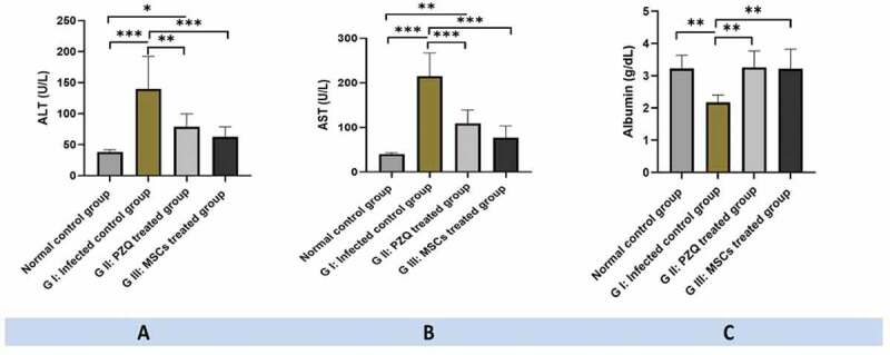
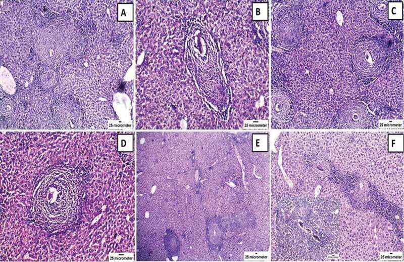
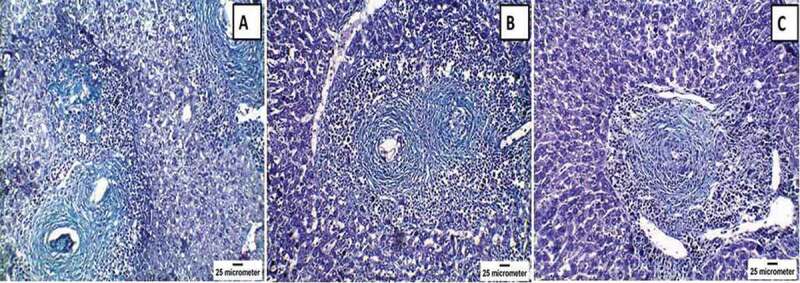
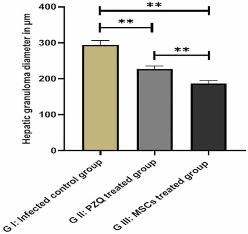
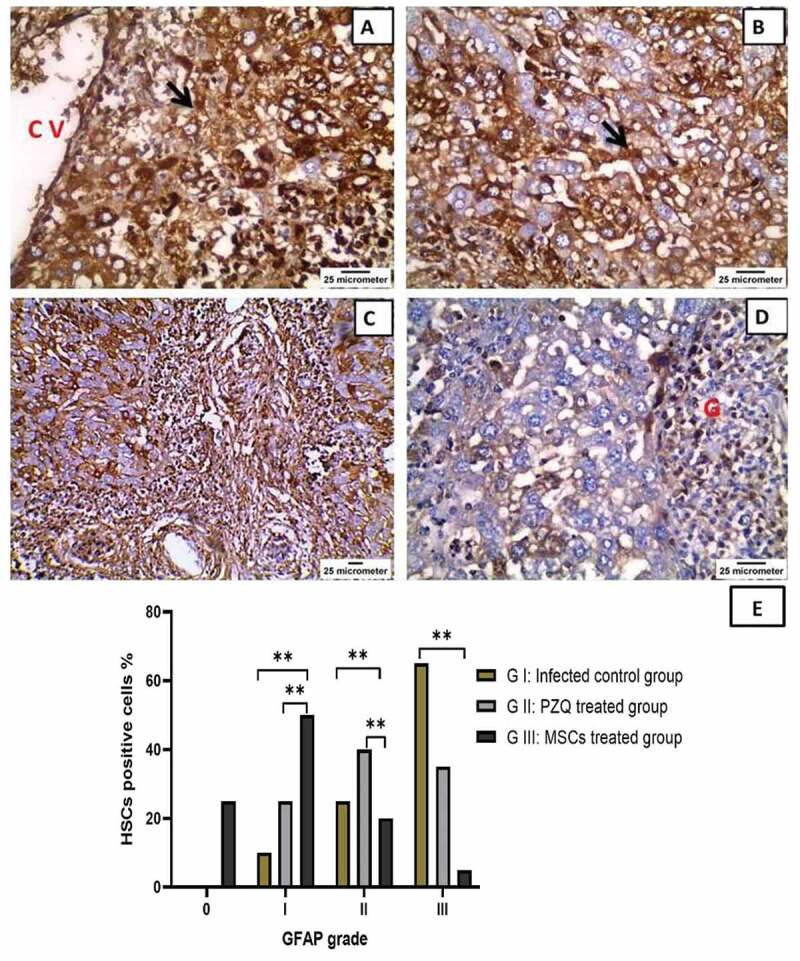
The objective of our study was to assess the effect of human umbilical cord blood (HUCB) mesenchymal stem cells (MSCs) transplantation on schistosomal hepatic fibrosis in mice. The study animals were divided into three groups. Group I is a control group, where the mice were infected with Schistosoma mansoni cercariae and remained untreated. The mice of the other two groups were infected and treated with either praziquantel (Group II) or HUCB-MSCs (Group III). Liver function tests, as well as histopathological evaluation of liver fibrosis using hematoxylin and eosin and Masson's trichrome stains, were performed. Additionally, an immunohistochemical study was carried out using anti-glial fibrillary acidic protein (GFAP) in hepatic stellate cells. Compared to the control group, the treated (praziquantel and MSCs) groups showed a substantial improvement, with a significant difference regarding the histopathological evaluation of liver fibrosis in the MSCs-treated group. In conclusion, MSCs could be a promising and efficient cell therapy for liver fibrosis.
Keywords: Liver fibrosis; glial fibrillary acidic protein; mesenchymal stem cells; praziquantel; schistosomiasis.
Conflict of interest statement
No potential conflict of interest was reported by the author(s).
Figures

Similar articles
-
Stem cell-derived exosomes as a potential therapy for schistosomal hepatic fibrosis in experimental animals.Pathog Glob Health. 2024 Jul;118(5):429-449. doi: 10.1080/20477724.2023.2240085. Epub 2023 Jul 30. Pathog Glob Health. 2024. PMID: 37519008 Free PMC article.
-
Cord blood-derived mesenchymal stem cells with hepatogenic differentiation potential ameliorate chronic liver affection in experimental models.Adv Clin Exp Med. 2018 Oct;27(10):1329-1339. doi: 10.17219/acem/70430. Adv Clin Exp Med. 2018. PMID: 30048056
-
Cotransplantation of human umbilical cord-derived mesenchymal stem cells and umbilical cord blood-derived CD34⁺ cells in a rabbit model of myocardial infarction.Mol Cell Biochem. 2014 Feb;387(1-2):91-100. doi: 10.1007/s11010-013-1874-5. Epub 2013 Oct 29. Mol Cell Biochem. 2014. PMID: 24166198
-
Prophylactic versus therapeutic role of the transplanted CD34+ Umbilical Cord Blood Stem Cells and Wharton Jelly Mesenchymal Stem Cells in early / acute hepatic S. mansoni granulomas reversal in mice; a novel approach.Exp Parasitol. 2020 Oct;217:107938. doi: 10.1016/j.exppara.2020.107938. Epub 2020 Aug 5. Exp Parasitol. 2020. PMID: 32768560 Review.
-
Immune microenvironment changes of liver cirrhosis: emerging role of mesenchymal stromal cells.Front Immunol. 2023 Jul 19;14:1204524. doi: 10.3389/fimmu.2023.1204524. eCollection 2023. Front Immunol. 2023. PMID: 37539053 Free PMC article. Review.
Cited by
-
Effectiveness and mechanisms of mesenchymal stem cell therapy in preclinical animal models of hepatic fibrosis: a systematic review and meta-analysis.Front Bioeng Biotechnol. 2024 Jul 22;12:1424253. doi: 10.3389/fbioe.2024.1424253. eCollection 2024. Front Bioeng Biotechnol. 2024. PMID: 39104627 Free PMC article.
-
Treatment of liver fibrosis: Past, current, and future.World J Hepatol. 2023 Jun 27;15(6):755-774. doi: 10.4254/wjh.v15.i6.755. World J Hepatol. 2023. PMID: 37397931 Free PMC article. Review.
-
New advances of NG2-expressing cell subset in marrow mesenchymal stem cells as novel therapeutic tools for liver fibrosis/cirrhosis.Stem Cell Res Ther. 2024 Jul 6;15(1):199. doi: 10.1186/s13287-024-03817-x. Stem Cell Res Ther. 2024. PMID: 38971781 Free PMC article.
-
Stem cell-derived exosomes as a potential therapy for schistosomal hepatic fibrosis in experimental animals.Pathog Glob Health. 2024 Jul;118(5):429-449. doi: 10.1080/20477724.2023.2240085. Epub 2023 Jul 30. Pathog Glob Health. 2024. PMID: 37519008 Free PMC article.
-
Protective effects and possible mechanisms of mesenchymal stem cells and mesenchymal stem cell-derived extracellular vesicles against kidney fibrosis in animal models: a systematic review and meta-analysis.Front Pharmacol. 2025 Jan 3;15:1511525. doi: 10.3389/fphar.2024.1511525. eCollection 2024. Front Pharmacol. 2025. PMID: 39830341 Free PMC article.
References
-
- Abou Rayia DM, Elmarhoumy SM, Ismail HH, et al. The outcomes of bone marrow stromal cell therapy in schistosomal hepatic fibrosis: an experimental study. J Egypt Soc Parasitol. 2017;47(3):633–642.
MeSH terms
Substances
LinkOut - more resources
Full Text Sources
Miscellaneous