. 2022 Jun;33(11-12):579-597.
doi: 10.1089/hum.2022.037.

Cell-Selective Adeno-Associated Virus-Mediated SCN1A Gene Regulation Therapy Rescues Mortality and Seizure Phenotypes in a Dravet Syndrome Mouse Model and Is Well Tolerated in Nonhuman Primates

Affiliations

Cell-Selective Adeno-Associated Virus-Mediated SCN1A Gene Regulation Therapy Rescues Mortality and Seizure Phenotypes in a Dravet Syndrome Mouse Model and Is Well Tolerated in Nonhuman Primates

Annie Tanenhaus et al. Hum Gene Ther. 2022 Jun.

Abstract

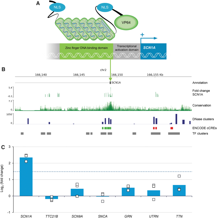
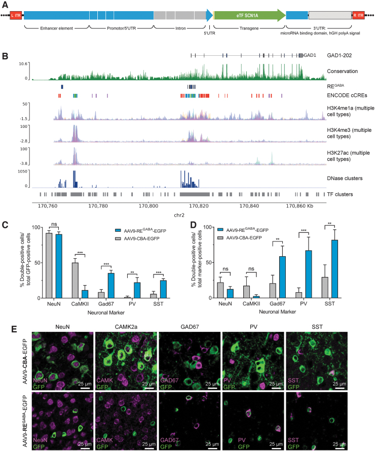
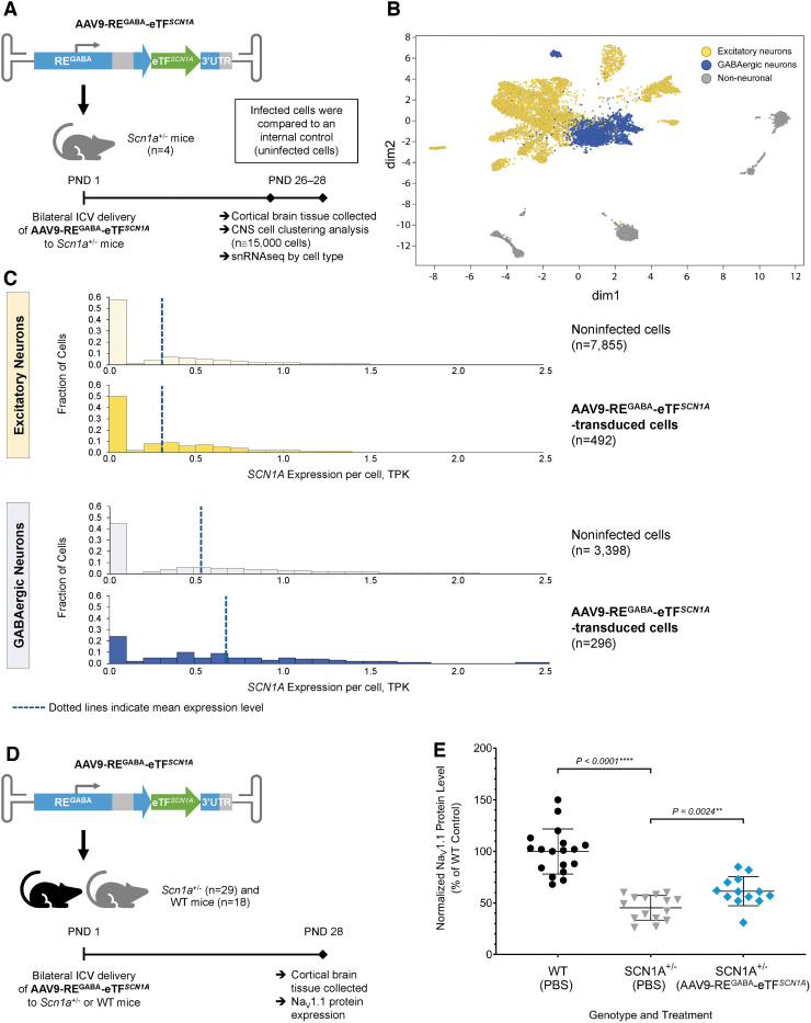
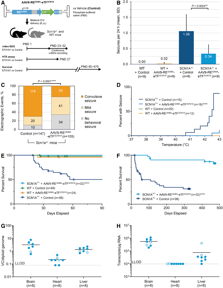
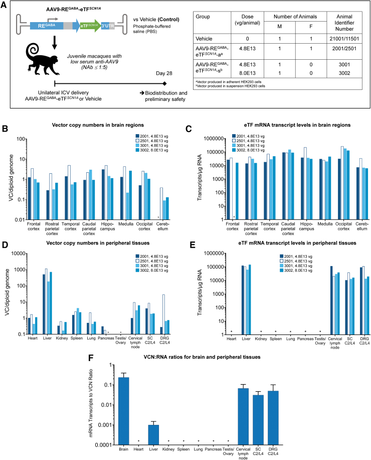
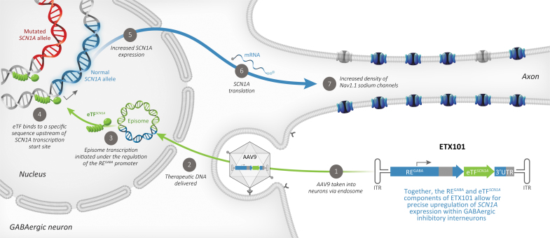
Dravet syndrome (DS) is a developmental and epileptic encephalopathy caused by monoallelic loss-of-function variants in the SCN1A gene. SCN1A encodes for the alpha subunit of the voltage-gated type I sodium channel (NaV1.1), the primary voltage-gated sodium channel responsible for generation of action potentials in GABAergic inhibitory interneurons. In these studies, we tested the efficacy of an adeno-associated virus serotype 9 (AAV9) SCN1A gene regulation therapy, AAV9-REGABA-eTFSCN1A, designed to target transgene expression to GABAergic inhibitory neurons and reduce off-target expression within excitatory cells, in the Scn1a+/- mouse model of DS. Biodistribution and preliminary safety were evaluated in nonhuman primates (NHPs). AAV9-REGABA-eTFSCN1A was engineered to upregulate SCN1A expression levels within GABAergic inhibitory interneurons to correct the underlying haploinsufficiency and circuit dysfunction. A single bilateral intracerebroventricular (ICV) injection of AAV9-REGABA-eTFSCN1A in Scn1a+/- postnatal day 1 mice led to increased SCN1A mRNA transcripts, specifically within GABAergic inhibitory interneurons, and NaV1.1 protein levels in the brain. This was associated with a significant decrease in the occurrence of spontaneous and hyperthermia-induced seizures, and prolonged survival for over a year. In NHPs, delivery of AAV9-REGABA-eTFSCN1A by unilateral ICV injection led to widespread vector biodistribution and transgene expression throughout the brain, including key structures involved in epilepsy and cognitive behaviors, such as hippocampus and cortex. AAV9-REGABA-eTFSCN1A was well tolerated, with no adverse events during administration, no detectable changes in clinical observations, no adverse findings in histopathology, and no dorsal root ganglion-related toxicity. Our results support the clinical development of AAV9-REGABA-eTFSCN1A (ETX101) as an effective and targeted disease-modifying approach to SCN1A+ DS.

Keywords: Dravet syndrome; SCN1A; channelopathy; encephalopathy; gene regulation therapy; preclinical models.


Conflict of interest statement

A.T., J.M., R.A., I.W.L., R.H., M.C., J. Li, S.P., M.C.V., M.M., A.B., and S.T. are current employees of and hold stock options in Encoded Therapeutics. A.Y., J.L., T.S., and T.C. are former employees of Encoded Therapeutics. J.A.K. serves as a consultant to Encoded Therapeutics, Praxis Precision Medicines, and NeuroCycle Therapeutics, and serves on the Scientific Advisory Boards of the Dravet Syndrome Foundation and FamilieSCN2A Foundation.

Figures

Figure 1.
Figure 1.
Structure of eTFSCN1A and its targeted activity at a unique and highly conserved 18-bp noncoding DNA sequence for gene-specific upregulation of SCN1A. (A) Schematic of eTFSCN1A structure. eTFSCN1A is composed of a polydactyl zinc finger DNA-binding domain, a short linker sequence, and a C-terminal VP64 transcriptional activation domain., Each of the six zinc finger domains interacts with a specific triplet nucleotide, conferring specific interaction with an 18-bp target sequence. NLS domains derived from simian vacuolating virus (SV40) and nucleoplasmin are included at the N-terminal and linker domains. (B) Genomic features of eTFSCN1A target site. Human genome track at the SCN1A locus. Tracks indicate human SCN1A gene annotation, chromosome 2 coordinates (hg38 genome assembly). FC SCN1A track shows target vector candidate screening data in HEK293cells. Mean Log2-FCs in SCN1A expression following transient transfection of eTFSCN1A candidates targeted against different genomic targets are plotted by target coordinates. The eTFSCN1A 18-bp target sequence, which induced the strongest upregulation of SCN1A within the screening window, is indicated by the green arrow. Sequence conservation track is indicated by 100-species base-wise conservation score (PhyloP). Regulatory element feature tracks are indicated (UCSC genome browser, ENCODE consortia,). DNase cluster track indicates DNase I hypersensitivity cluster score; ENCODE cCREs track marks candidate cis-regulatory element regions (green: PLS, red: dELS); TF clusters track marks transcription factor binding peak clusters. (C) eTFSCN1A upregulates SCN1A in a gene-specific manner in vitro. Change in endogenous gene expression in HEK293 cells is reported in the eTFSCN1A transfected condition relative to control condition for each replicate. Bars represent mean Log2-FC for n = 3 biological replicates (n = 2 for PAPBC1), dots indicate individual replicate measurements. Dotted line indicates Log2FC = 1.5. bp, base-pair; cCREs, candidate cis-regulatory elements; dELS, distal enhancer-like signature; FC, fold change; NLS, nuclear localization signal; PLS, promoter-like signature; TF, transcription factor.
Figure 2.
Figure 2.
The REGABA regulatory element in AAV9-REGABA-eTFSCN1A selectively targets GABAergic interneurons in vivo. (A) REGABA construct design. REGABA comprised upstream and 5′UTR sequence components, and a downstream 3′UTR sequence. The upstream regulatory sequence is derived from human genomic sequence elements: an enhancer element located ∼50 kb upstream of the GAD1 gene locus, and proximal promoter and 5′UTR element sequence derived from the human GAD1 gene locus. The promoter is composed of genomic sequence segments in the proximal promoter and 5′UTR of the hGAD1 gene. To enhance gene expression and to capture additional regulatory features derived from intronic elements, this promoter also incorporates a truncated intron derived from the first intron of the GAD1 gene, which includes sequences flanking the splice donor and acceptor sites, and internal intronic sequences that overlap high-conservation predicted regulatory sequences. The 3′UTR element includes a collection of eight cognate target motifs derived from excitatory neuron-enriched miRNAs miR128 and mir221,, and a hGH-pA. (B) Genomic features of REGABA source sequence. Human genome track at the human GAD1 locus. Tracks indicate GAD1 gene annotation, chromosome 2 coordinates (hg38 genome assembly). Sequence conservation (PhyloP,), and ENCODE regulatory element feature tracks are indicated, as well as additional composite epigenetic marker tracks for histone markers H3K4me1, H3K4me3, and H3K27ac, which can signal active enhancer and promoter regions (UCSC genome browser, ENCODE consortia). (C–E) Expression of EGFP in vivo 27 days post-ICV injection of AAV9-EGFP vectors driven by CBA or REGABA promoters in PND1 mice. Animals (n = 4/group) were administered 2.0E10 vg per animal of AAV-CBA-EGFP or AAV-REGABA-EGFP on PND1 by bilateral ICV injection. On PND28, brain sections were analyzed by IHC for the presence of GFP and neuron-specific markers. Quantitation of colocalization between neuron-specific markersa and EGFP: (C) among GFP-positive cells and (D) compared to the total number of neuron-marker positive cells; **p < 0.01; ***p < 0.001; ns, p ≥ 0.05 (unpaired t-tests, two-stage step-up FDR = 1%; data shown are means and error bars represent SD). (E) Representative images demonstrating colocalization of EGFP driven by each vector with each of the neuronal markers evaluated. aNeuron-specific markers: NeuN (neurons); CAMK2a (glutamatergic neurons); GAD67 (GABAergic interneurons); PV (parvalbumin-positive interneurons); SST (somatostatin-positive interneurons). AAV, adeno-associated virus; CBA, chicken β-actin promoter; chr2, chromosome 2; EGFP, enhanced green fluorescent protein; eTF, engineered transcription factor; hGH-pA, human growth hormone-derived poly-adenylation signal; IHC, immunohistochemistry; ICV, intracerebroventricular; ITR, inverted terminal repeat; ns, not significant; PND, postnatal day; REGABA, GABAergic cell-selective regulatory element; SD, standard deviation; UTR, untranslated region; vg, vector genomes.
Figure 3.
Figure 3.
AAV9-REGABA-eTFSCN1A upregulates SCN1A within GABAergic inhibitory interneurons in vivo. (A) Experimental design to evaluate SCN1A expression in excitatory and GABAergic inhibitory neurons. Four Scn1a+/− mice were treated with 1.7E10 vg per animal of AAV9-REGABA-eTFSCN1A on PND1 via bilateral ICV injection. On PND28, cortical brain tissue was collected, and clustering analysis was performed to classify over 15,000 cells into excitatory and inhibitory cell types based on transcriptome profiles. (B) Clustering of CNS cell types in cortical brain tissue. Gene expression visualization using tSNE to compute a low-dimensional representation of snRNAseq data; dim2 and dim1 are the outputs of tSNE, the 2D representation of the gene expression profiles of each cell. Yellow dots denote excitatory neurons; blue dots denote GABAergic inhibitory neurons; gray dots denote non-neuronal cells. (C) SCN1A mRNA transcript levels in excitatory and GABAergic inhibitory neurons. AAV9-REGABA-eTFSCN1A upregulates SCN1A mRNA specifically in GABAergic inhibitory neurons, with no upregulation in excitatory neurons. Dotted lines indicate mean SCN1A TPK per cell. (D) Experimental design to determine NaV1.1 protein expression. One-day-old (PND1) Scn1a+/− mice were administered PBS (n = 16) or 5.1E10 vg per animal AAV9-REGABA-eTFSCN1A (n = 13) and 18 WT littermates were administered PBS via a bilateral ICV injection. Animals were sacrificed on PND28 and brain tissue was evaluated by MSD electrochemiluminescence-based sandwich immunoassay. (E) NaV1.1 protein expression. Membrane-associated NaV1.1 protein levels from PBS-treated WT (6 males/12 females; black circle) or Scn1a+/− mice (7 males/9 females; gray triangle) and AAV9-REGABA-eTFSCN1A-treated Scn1a+/− mice (9 males/4 females; blue diamond). Mean levels ± SD are indicated; ****p < 0.0001 and **p = 0.0024, Mann–Whitney test. CNS, central nervous system; dim, dimension; MSD, Meso Scale Discovery; NaV1.1, voltage-gated type I sodium channel; PBS, phosphate-buffered saline; snRNAseq, single-nucleus RNA sequencing analysis; TPK, transcripts per 1000 total transcripts; tSNE, t-distributed stochastic neighbor embedding; WT, wild-type.
Figure 4.
Figure 4.
AAV9-REGABA-eTFSCN1A reduces frequency and severity of spontaneous seizures, protects against febrile seizures, and demonstrates durable survival efficacy and persistent activity for up to 470 days postdosing in Scn1a+/− mice. (A) Study design. AAV9-REGABA-eTFSCN1A or vehicle alone (PBS) were administered by bilateral ICV injection (3 μL/hemisphere) to heterozygous Scn1a+/− or WT littermates at PND1. Separate studies assessed electrographic seizure monitoring of spontaneous seizures (following doses of 1.2–5.4E11 vg/animala vs. vehicle); susceptibility to HTS (3.7E10 vg/animala vs. vehicle); and survival and long-term persistence of AAV9-REGABA-eTFSCN1A (3.7E10 vg/animala vs. vehicle). At 470 days, the animals were sacrificed, and brain, heart, and liver tissues were evaluated for AAV9-REGABA-eTFSCN1A VCN by ddPCR and eTF transgene mRNA transcript levels by RT-ddPCR. (B) EEG seizure frequency. Mice treated with AAV9-REGABA-eTFSCN1A experienced a 68% reduction in the mean daily generalized seizure frequency per animal compared with Scn1a+/− controls (**p = 0.0024, unpaired t-test). No changes in seizure frequency were detected in WT mice treated with AAV9-REGABA-eTFSCN1A. (C) EEG seizure severity. Video characterization of all recorded electrographic seizure events revealed that treatment of Scn1a+/− mice with AAV9-REGABA-eTFSCN1A significantly reduced convulsive (tonic-clonic) seizures compared with Scn1a+/− mice treated with vehicle (****p < 0.0001; chi-square test). Most events detected in AAV9-REGABA-eTFSCN1A-treated Scn1a+/– mice were mild (characterized by mild movements and/or head twitching, but without convulsions) or nonbehavioral seizures. (D) HTS assay. Percentage of AAV9-REGABA-eTFSCN1A PND1-treated Scn1a+/− mice experiencing seizures at a given temperature at PND27. WT groups overlap along the zero line (green and orange lines; only the orange trace is shown). p-Values calculated using a Log-rank test, ****p < 0.0001. (E) Ninety-day survival. The 90-day survival rate in AAV9-REGABA-eTFSCN1A-treated Scn1a+/– mice was 100% compared with 50% in control-treated Scn1a+/− mice (****p < 0.0001, Log-rank test). No difference in survival was observed between WT mice ± AAV9-REGABA-eTFSCN1A and Scn1a+/− mice + AAV9-REGABA-eTFSCN1A. (F) Long-term Survival. Long-term follow-up showed that survival benefit of AAV9-REGABA-eTFSCN1A was sustained over ∼470 days after dosing (****p < 0.0001, Log-rank test). Diamond indicates humane endpoint or end of study euthanasia at approximately day 330 for vehicle and day 470 for AAV9-REGABA-eTFSCN1A-treated animals. (G) VCN biodistribution in Scn1a+/− mice administered AAV9-REGABA-eTFSCN1A. At 470 days, the animals were sacrificed, and brain, heart, and liver tissues were evaluated for AAV9-REGABA-eTFSCN1A VCN by ddPCR and eTFSCN1A transgene mRNA transcripts levels by RT-ddPCR. (H) eTFSCN1A mRNA expression levels in Scn1a+/− mice administered AAV9-REGABA-eTFSCN1A. eTFSCN1A mRNA transcripts per microgram of RNA analyzed. Seven of eight animals had no measurable eTFSCN1A mRNA transcripts in the heart. Open circle indicates female gender. Dotted line indicates the limit of detection in both assays. aDoses in the EEG study were determined by a qPCR titering method while the HTS and survival studies were titered via ddPCR; thus the doses used across studies are not directly comparable. EEG, electroencephalography; HTS, hyperthermia-induced seizure; LLOD, lower limit of detection; qPCR, quantitative polymerase chain reaction; RT-ddPCR, reverse transcription digital droplet polymerase chain reaction; VCN, vector copy number.
Figure 5.
Figure 5.
Unilateral ICV delivery of AAV9-REGABA-eTFSCN1A to NHPs leads to widespread vector biodistribution and increased eTFSCN1A mRNA transcript levels in the brain with low off-target vector expression in peripheral tissues of NHPs. (A) Study design. AAV9-REGABA-eTFSCN1A (4.8–8.0 E13 vg/animal) or vehicle alone (PBS) was administered by unilateral ICV injection to four juvenile cynomolgus macaques (3M and 1F). All animals were sacrificed 28 ± 2 days after injection. Biodistribution of AAV9-REGABA-eTFSCN1A vector copies and expression of transgene mRNA were measured in target neuronal and peripheral tissues using ddPCR (B–E). (B) AAV9-REGABA-eTFSCN1A vector biodistribution in brain regions. (C) eTFSCN1A mRNA expression levels in brain regions. (D) AAV9-REGABA-eTFSCN1A vector biodistribution and (E) eTFSCN1A mRNA expression levels in peripheral tissues. (F) VCN:RNA ratios for brain and peripheral tissues (N = 4, mean values; error bars indicate SD). Starred organs (*) indicate VCN or RNA levels below the limit of detection of the assay. DRG, dorsal root ganglia; NAb, neutralizing antibodies; NHP, nonhuman primate; SC, spinal cord.
Figure 6.
Figure 6.
Mechanism of action of AAV9-REGABA-eTFSCN1A (ETX101), a cell-selective AAV-mediated SCN1A gene regulation therapy that upregulates expression of WT SCN1A selectively in GABAergic inhibitory interneurons to compensate for the loss-of-function mutant alleles in individuals with DS. As previously described, AAV vector enters the cell by endocytosis in a receptor-mediated manner. Vector escapes from the endosome, enters the nucleus through the nuclear pore complex, and predominantly exists as a nonreplicating episome. Episome transcription is then initiated under the regulation of the REGABA promoter to produce eTFSCN1A preferably in GABAergic neurons. eTFSCN1A binds to a conserved regulatory region upstream of the SCN1A transcription start site, promoting increased SCN1A expression and protein translation, thereby increasing the density of membrane-associated NaV1.1 sodium channels and restoring function. While eTFSCN1A also binds the mutated SCN1A allele, it does not produce any stable protein capable of functioning at the neuronal membrane. This approach leverages natural patterns of gene expression to increase production of NaV1.1 protein and restore inhibitory function while minimizing potential off target effects. DS, Dravet syndrome; RE, regulatory element.

References

    1. Dravet C, Guerrini R. Dravet Syndrome. Topics in Epilepsy, 3rd ed. Paris: John Libbey Eurotext, 2011.
    1. Nabbout R, Chemaly N, Chipaux M, et al. . Encephalopathy in children with Dravet syndrome is not a pure consequence of epilepsy. Orphanet J Rare Dis 2013;8:176. - PMC - PubMed
    1. Brunklaus A, Ellis R, Reavey E, et al. . Prognostic, clinical and demographic features in SCN1A mutation-positive Dravet syndrome. Brain 2012;135(Pt 8):2329–2336. - PubMed
    1. Gataullina S, Dulac O. From genotype to phenotype in Dravet disease. Seizure 2017;44:58–64. - PubMed
    1. Wirrell EC, Laux L, Donner E, et al. . Optimizing the diagnosis and management of Dravet syndrome: recommendations from a North American Consensus Panel. Pediatr Neurol 2017;68:18–34.e3. - PubMed

Publication types

Substances

Supplementary concepts

LinkOut - more resources