. 2022 Mar 31:(181):10.3791/63666.
doi: 10.3791/63666.

Laser Microdissection for Species-Agnostic Single-Tissue Applications

Affiliations

Laser Microdissection for Species-Agnostic Single-Tissue Applications

Alyssa Woronik et al. J Vis Exp. .

Abstract

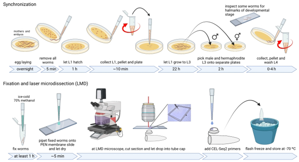
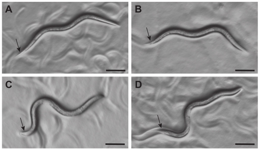
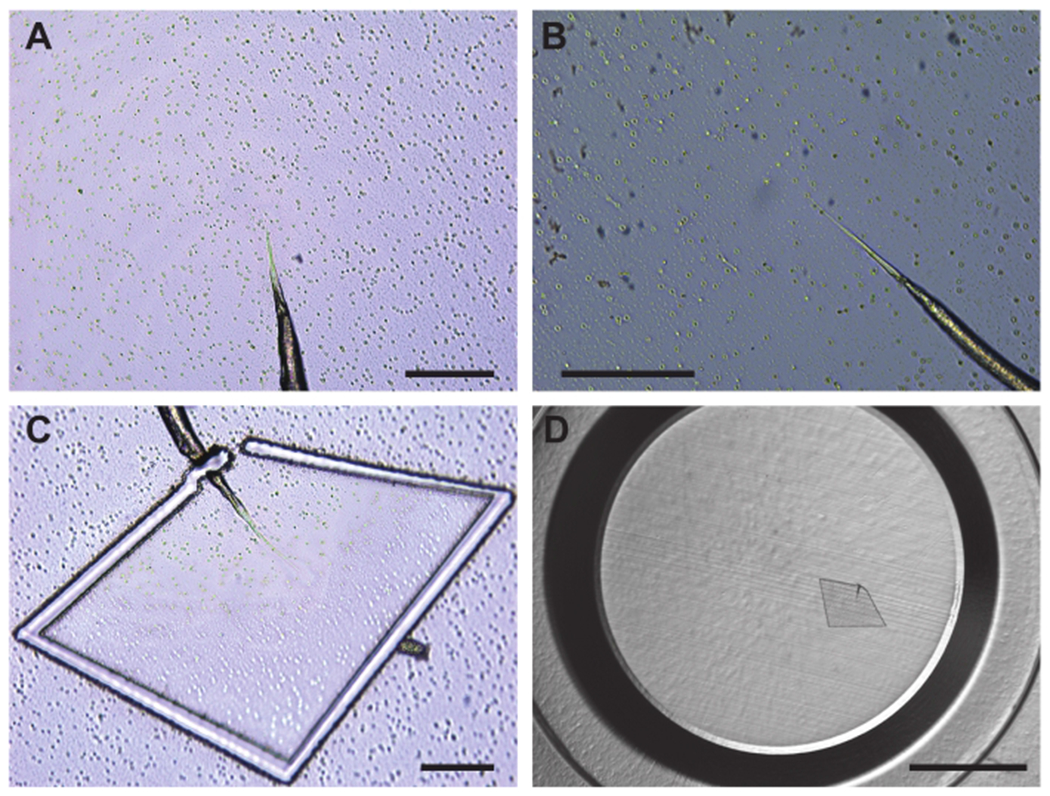
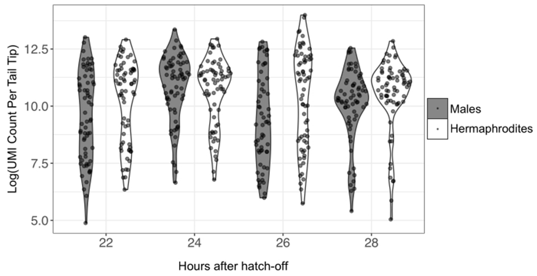
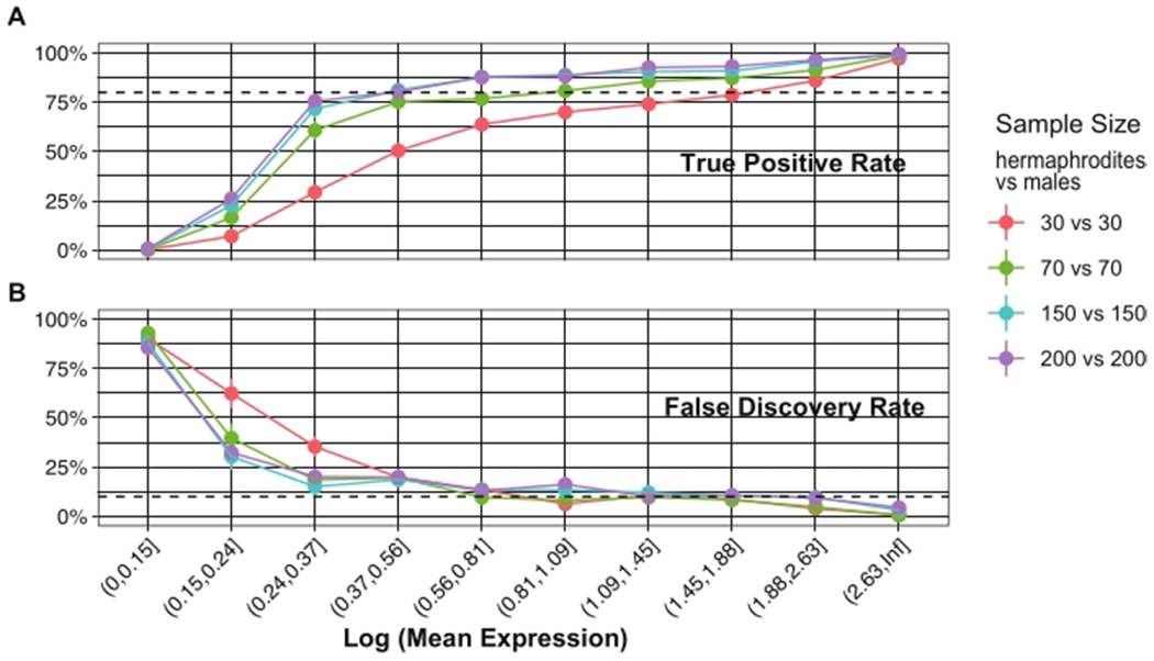
Single-cell methodologies have revolutionized the analysis of the transcriptomes of specific cell types. However, they often require species-specific genetic "toolkits," such as promoters driving tissue-specific expression of fluorescent proteins. Further, protocols that disrupt tissues to isolate individual cells remove cells from their native environment (e.g., signaling from neighbors) and may result in stress responses or other differences from native gene expression states. In the present protocol, laser microdissection (LMD) is optimized to isolate individual nematode tail tips for the study of gene expression during male tail tip morphogenesis. LMD allows the isolation of a portion of the animal without the need for cellular disruption or species-specific toolkits and is thus applicable to any species. Subsequently, single-cell RNA-seq library preparation protocols such as CEL-Seq2 can be applied to LMD-isolated single tissues and analyzed using standard pipelines, given that a well-annotated genome or transcriptome is available for the species. Such data can be used to establish how conserved or different the transcriptomes are that underlie the development of that tissue in different species. Limitations include the ability to cut out the tissue of interest and the sample size. A power analysis shows that as few as 70 tail tips per condition are required for 80% power. Tight synchronization of development is needed to obtain this number of animals at the same developmental stage. Thus, a method to synchronize animals at 1 h intervals is also described.


Conflict of interest statement

Disclosures

All authors declare that they have no conflicts of interest.

Figures

Figure 1:
Figure 1:. Procedure overview for synchronization of Caenorhabditis elegans with the hatch-off method and laser microdissection of tail tips.
Abbreviations: L1-L4 = larval stages 1 to 4; PEN = polyethylene naphthalate; LMD = laser microdissection. Please click here to view a larger version of this figure.
Figure 2:
Figure 2:. Appearance of C. elegans L3 hermaphrodites and males under a dissection microscope.
Hermaphrodites (A, B) and males (C, D) at 21-23 h after hatch can be distinguished under a dissection microscope (~50x magnification) by the morphology of their tails (arrows). The tail of hermaphrodites is narrow, while that of males is swollen and appears clear. Scale bars = 0.1 mm. Please click here to view a larger version of this figure.
Figure 3:
Figure 3:. Appearance of the PEN membrane slide structure and worm tail.
Focus is correct for dissection of the tissue viewed with the 20x (A) and 40x (B) lens at the microscope. (C) Dissected tail and partially cut out PEN membrane. After closing the gap in the cut, the membrane piece will drop into the tube cap below the slide. (D) Tube cap with a PEN membrane section containing a dissected tail tip. Scale bars = 0.1 mm (A-C), 1 mm (D). Please click here to view a larger version of this figure.
Figure 4:
Figure 4:. Natural log-transformed UMI counts per individual tail tip for different time points and sexes.
RNA from individual tails was prepared for sequencing using the CEL-Seq2 method; 557 tails were sequenced in total, with 59-78 tails per sex and time point. Extremely low and high UMI outliers would be removed from the data before analysis. Abbreviation: UMI = unique molecular identifier. Please click here to view a larger version of this figure.
Figure 5:
Figure 5:. Results of an a posteriori power analysis using simulations with powsimR.
The powsimR software determines the number of independent samples required to detect DE genes at various expression levels. Genes are binned by mean expression transformed as the natural log of UMI counts. (A) Power (TPR) to detect DE genes between two conditions (here, male vs hermaphrodite) for four different simulations (different colored graphs) incorporating different sample sizes (numbers of individual tail-tips) per condition. Dashed line indicates 80% TPR. (B) FDR in the same four simulations as in (A), dashed line indicating 10% FDR. The graphs show that a sample size of 70 tail tips (green) per condition is sufficient for detecting DE genes, except for genes with very low expression levels. That is, the power and false discovery rate for such genes cannot be greatly improved by increasing the sample size beyond 70. Abbreviations: DE = differentially expressed; UMI = unique molecular identifier; TPR = true positive rate; FDR = false discovery rate. Please click here to view a larger version of this figure.

References

    1. Haag ES, Fitch DHA, Delattre M From “the worm” to “the worms” and back again: the evolutionary developmental biology of nematodes. Genetics. 210 (2), 397–433 (2018). - PMC - PubMed
    1. Sommer RJ, Bumbarger DJ Nematode model systems in evolution and development. Wiley Interdisciplinary Reviews: Developmental Biology. 1 (3), 389–400 (2012). - PubMed
    1. WormBase. https://wormbase.org/#012-3-6 (2022).
    1. WormAtlas. https://www.wormatlas.org (2022).
    1. WormBook. http://wormbook.org (2022).

Publication types

LinkOut - more resources