Review
. 2022 Aug 2;28(15):3196-3206.
doi: 10.1158/1078-0432.CCR-21-1356.

Immunotherapy of Neuroblastoma: Facts and Hopes

Affiliations
Review

Immunotherapy of Neuroblastoma: Facts and Hopes

John Anderson et al. Clin Cancer Res. .

Abstract

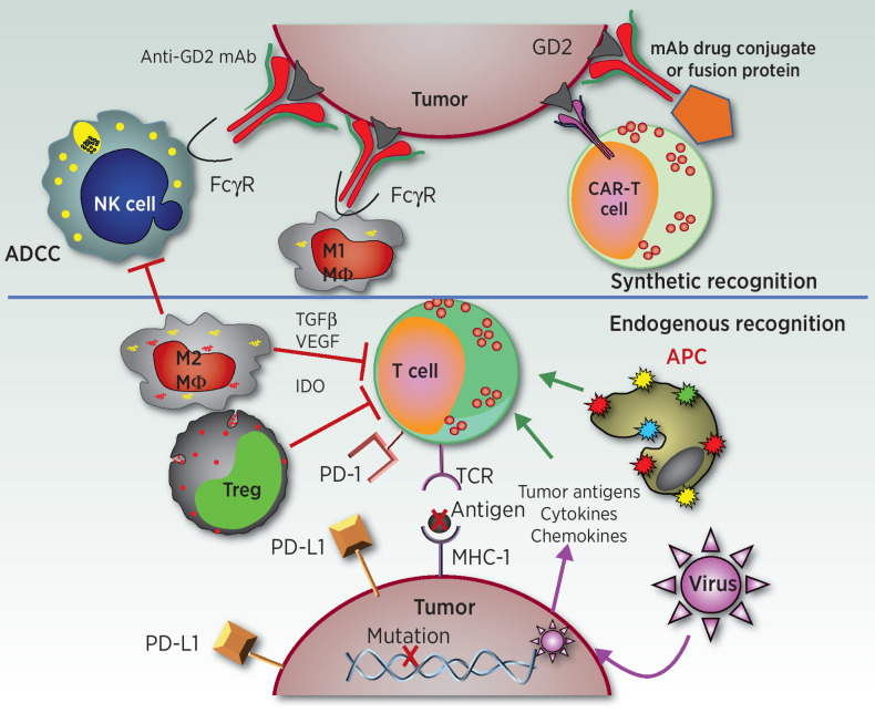
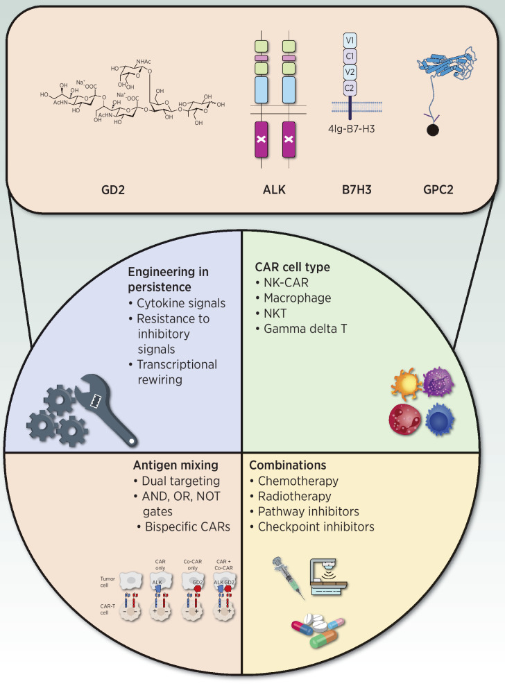
While the adoption of multimodal therapy including surgery, radiation, and aggressive combination chemotherapy has improved outcomes for many children with high-risk neuroblastoma, we appear to have reached a plateau in what can be achieved with cytotoxic therapies alone. Most children with cancer, including high-risk neuroblastoma, do not benefit from treatment with immune checkpoint inhibitors (ICI) that have revolutionized the treatment of many highly immunogenic adult solid tumors. This likely reflects the low tumor mutation burden as well as the downregulated MHC-I that characterizes most high-risk neuroblastomas. For these reasons, neuroblastoma represents an immunotherapeutic challenge that may be a model for the creation of effective immunotherapy for other "cold" tumors in children and adults that do not respond to ICI. The identification of strong expression of the disialoganglioside GD2 on the surface of nearly all neuroblastoma cells provided a target for immune recognition by anti-GD2 mAbs that recruit Fc receptor-expressing innate immune cells that mediate cytotoxicity or phagocytosis. Adoption of anti-GD2 antibodies into both upfront and relapse treatment protocols has dramatically increased survival rates and altered the landscape for children with high-risk neuroblastoma. This review describes how these approaches have been expanded to additional combinations and forms of immunotherapy that have already demonstrated clear clinical benefit. We also describe the efforts to identify additional immune targets for neuroblastoma. Finally, we summarize newer approaches being pursued that may well help both innate and adaptive immune cells, endogenous or genetically engineered, to more effectively destroy neuroblastoma cells, to better induce complete remission and prevent recurrence.


Figures

Figure 1. Endogenous and synthetic recognition involved in immunotherapies for neuroblastoma. This simplified schematic shows some of the relationships between immune cells, molecules, and cancer cells involved in current and developing immunotherapies for neuroblastoma. The synthetic recognition pathways are shown above the horizontal line, and are all shown here as mediated via mAb-induced tumor recognition. The mAb-based tumor-recognition components, shown at the top, include an intact anti-GD2 mAb (at top left) binding to GD2 on the tumor engaging the Fcγ receptor (FcγR) on the NK cell or on the M1 macrophage (MΦ) to activate ADCC. At the top right is that same anti-GD2 mAb, now carrying a “payload.” This payload can be a drug, as an ADC, an immune activator as in a fusion protein, such as an IL2-linked immunocytokine, a radionuclide, or a toxin. To its left is a CAR-T cell that utilizes the ScFv of the anti-GD2 mAb to provide anti-GD2 recognition for the genetically modified T cell. Below the horizontal line are the pathways involved in endogenous recognition, with a central role given to effector T cells. At the bottom of the endogenous T cell is its T-cell antigen receptor (TCR), which on clonally derived T cells can recognize tumor-associated peptides presented by the MHC molecules on the tumor surface. This recognition and T-cell activation can induce effector functions, that include cytokine release, activation of innate immune antitumor cells, and direct T cell–mediated tumor cell lysis. Those tumor associated peptides can be mutation-driven neoantigens (shown here) or germline controlled proteins that have restricted expression to tumor cells, with little or no expression on normal postnatal tissues. To the left of the T cell are endogenous cells that can interfere with T-cell function. One such inhibitory cell is an M2 macrophage (MΦ), that can interfere with antitumor immunotherapy via many pathways, including release of TGF β, VEGF and indoleamine 2,3-dioxygenase (IDO). Other myeloid elements, like myeloid-derived suppressor cells (not shown) can also interfere with effector immune function. Regulatory T cells (Treg) are normally FoxP3+ CD4+ T cells that can directly kill or inhibit the functions of effector T cells. These inhibitory cells can also interfere with NK-cell function (not shown). To the right of the T cell is an antigen-presenting cell (APC), normally a dendritic cell, that picks up and processes tumor antigens and then presents them to T cells to induce an endogenous adaptive immune response. Cytokines and chemokines can help recruit immune cells into the tumor. Certain oncolytic tumor viruses are being injected in some trials to infect the tumor, release more chemokines, and recruit additional immune cells to the tumor microenvironment. The immunosuppressive PD-L1 ligand, is one of several checkpoint molecules expressed by tumor cells (and shown here). PD-L1 activates the immune-inhibitory PD-1 receptor on the T cell (shown) and some NK cells (not shown). Not depicted is how anti-PD1 or anti-PD-L1 mAbs (forms of immune checkpoint blockade) can block these inhibitory interactions, enabling T and NK cells functionality in the suppressive tumor microenvironment.
Figure 1.
Endogenous and synthetic recognition involved in immunotherapies for neuroblastoma. This simplified schematic shows some of the relationships between immune cells, molecules, and cancer cells involved in current and developing immunotherapies for neuroblastoma. The synthetic recognition pathways are shown above the horizontal line, and are all shown here as mediated via mAb-induced tumor recognition. The mAb-based tumor-recognition components, shown at the top, include an intact anti-GD2 mAb (at top left) binding to GD2 on the tumor engaging the Fcγ receptor (FcγR) on the NK cell or on the M1 macrophage (MΦ) to activate ADCC. At the top right is that same anti-GD2 mAb, now carrying a “payload.” This payload can be a drug, as an ADC; an immune activator as in a fusion protein, such as an IL2-linked immunocytokine; a radionuclide; or a toxin. To its left is a CAR-T cell that utilizes the ScFv of the anti-GD2 mAb to provide anti-GD2 recognition for the genetically modified T cell. Below the horizontal line are the pathways involved in endogenous recognition, with a central role given to effector T cells. At the bottom of the endogenous T cell is its T-cell antigen receptor (TCR), which on clonally derived T cells can recognize tumor-associated peptides presented by the MHC molecules on the tumor surface. This recognition and T-cell activation can induce effector functions, including cytokine release, activation of innate immune antitumor cells, and direct T cell–mediated tumor cell lysis. Those tumor-associated peptides can be mutation-driven neoantigens (shown here) or germline controlled proteins that have restricted expression to tumor cells, with little or no expression on normal postnatal tissues. To the left of the T cell are endogenous cells that can interfere with T-cell function. One such inhibitory cell is an M2 macrophage (MΦ), which can interfere with antitumor immunotherapy via many pathways, including release of TGFβ, VEGF, and indoleamine 2,3-dioxygenase (IDO). Other myeloid elements, such as myeloid-derived suppressor cells (not shown), can also interfere with effector immune function. Regulatory T cells (Treg) are normally FoxP3+ CD4+ T cells that can directly kill or inhibit the functions of effector T cells. These inhibitory cells can also interfere with NK-cell function (not shown). To the right of the T cell is an antigen-presenting cell (APC), normally a dendritic cell, that picks up and processes tumor antigens and then presents them to T cells to induce an endogenous adaptive immune response. Cytokines and chemokines can help recruit immune cells into the tumor. Certain oncolytic tumor viruses are being injected in some trials to infect the tumor, release more chemokines, and recruit additional immune cells to the tumor microenvironment. The immunosuppressive PD-L1 ligand is one of several checkpoint molecules expressed by tumor cells (and shown here). PD-L1 activates the immune-inhibitory PD-1 receptor on the T cell (shown) and some NK cells (not shown). Not depicted is how anti-PD1 or anti–PD-L1 mAbs (forms of immune checkpoint blockade) can block these inhibitory interactions, enabling T and NK cell functionality in the suppressive tumor microenvironment.
Figure 2. Enhancing CAR-T cell sensitivity and specificity. Schematic of technological solutions to enhance CAR-T cell sensitivity and specificity. Representative neuroblastoma tumor antigens that are currently under clinical and preclinical evaluation are indicated at the top. At the bottom are the categories of approaches for enhancing functionality of CAR-T cells that are relevant to neuroblastoma.
Figure 2.
Enhancing CAR-T cell sensitivity and specificity. Schematic of technologic solutions to enhance CAR-T cell sensitivity and specificity. Representative neuroblastoma tumor antigens that are currently under clinical and preclinical evaluation are indicated at the top. At the bottom are the categories of approaches for enhancing functionality of CAR-T cells that are relevant to neuroblastoma.

References

    1. Postow MA, Chesney J, Pavlick AC, Robert C, Grossmann K, McDermott D, et al. . Nivolumab and ipilimumab versus ipilimumab in untreated melanoma. N Engl J Med 2015;372:2006–17. - PMC - PubMed
    1. Reck M, Rodriguez-Abreu D, Robinson AG, Hui R, Csoszi T, Fulop A, et al. . Pembrolizumab versus chemotherapy for PD-L1-positive non-small-cell lung cancer. N Engl J Med 2016;375:1823–33. - PubMed
    1. Maude SL, Laetsch TW, Buechner J, Rives S, Boyer M, Bittencourt H, et al. . Tisagenlecleucel in children and young adults with B-cell lymphoblastic leukemia. N Engl J Med 2018;378:439–48. - PMC - PubMed
    1. Neelapu SS, Locke FL, Bartlett NL, Lekakis LJ, Miklos DB, Jacobson CA, et al. . Axicabtagene ciloleucel CAR T-cell therapy in refractory large B-cell lymphoma. N Engl J Med 2017;377:2531–44. - PMC - PubMed
    1. Merchant MS, Wright M, Baird K, Wexler LH, Rodriguez-Galindo C, Bernstein D, et al. . Phase I clinical trial of Ipilimumab in pediatric patients with advanced solid tumors. Clin Cancer Res 2016;22:1364–70. - PMC - PubMed

Publication types