. 2022 Jun 1;322(6):R609-R619.
doi: 10.1152/ajpregu.00027.2022. Epub 2022 Apr 19.

Tilapia prolactin cells are thermosensitive osmoreceptors

Affiliations

Tilapia prolactin cells are thermosensitive osmoreceptors

Daniel W Woo et al. Am J Physiol Regul Integr Comp Physiol. .

Abstract

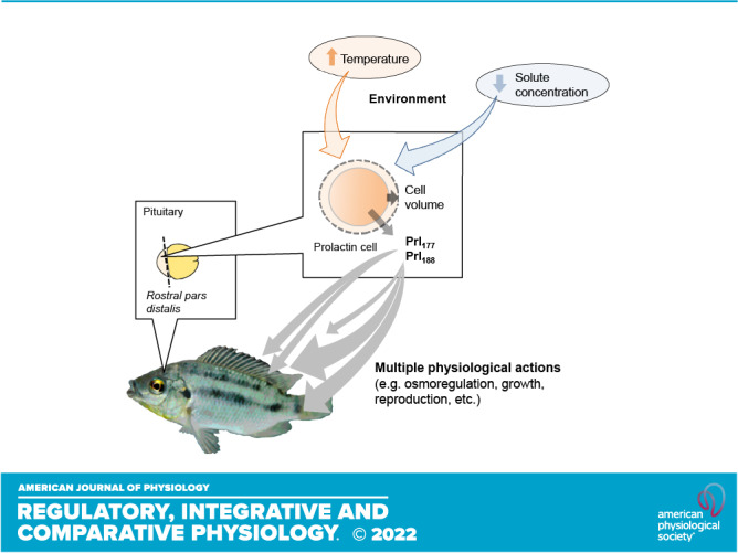
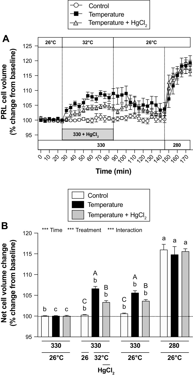
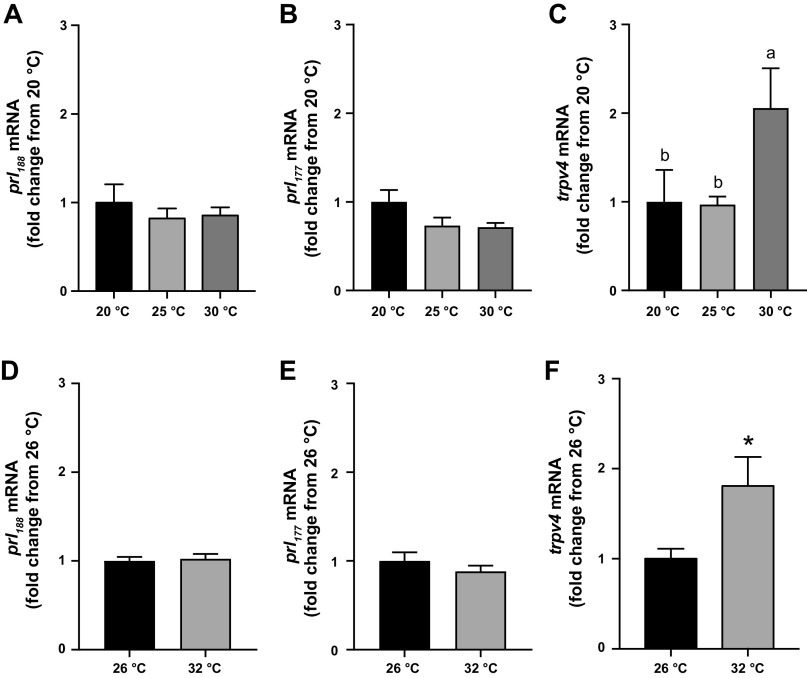
Prolactin (PRL) cells within the rostral pars distalis (RPD) of euryhaline and eurythermal Mozambique tilapia, Oreochromis mossambicus, rapidly respond to a hyposmotic stimulus by releasing two distinct PRL isoforms, PRL188 and PRL177. Here, we describe how environmentally relevant temperature changes affected mRNA levels of prl188 and prl177 and the release of immunoreactive prolactins from RPDs and dispersed PRL cells. When applied under isosmotic conditions (330 mosmol/kgH2O), a 6°C rise in temperature stimulated the release of PRL188 and PRL177 from both RPDs and dispersed PRL cells under perifusion. When exposed to this same change in temperature, ∼50% of dispersed PRL cells gradually increased in volume by ∼8%, a response partially inhibited by the water channel blocker, mercuric chloride. Following their response to increased temperature, PRL cells remained responsive to a hyposmotic stimulus (280 mosmol/kgH2O). The mRNA expression of transient potential vanilloid 4, a Ca2+-channel involved in hyposmotically induced PRL release, was elevated in response to a rise in temperature in dispersed PRL cells and RPDs at 6 and 24 h, respectively; prl188 and prl177 mRNAs were unaffected. Our findings indicate that thermosensitive PRL release is mediated, at least partially, through a cell-volume-dependent pathway similar to how osmoreceptive PRL release is achieved.

Keywords: fish; osmoreceptor; prolactin; thermoreceptor; thermosensitivity.


Conflict of interest statement

No conflicts of interest, financial or otherwise, are declared by the authors.

Figures

None
Graphical abstract
Figure 1.
Figure 1.
Diagram summarizing the experimental setup used for determining changes in PRL cell volume in responses to a rise in temperature. PRL, prolactin.
Figure 2.
Figure 2.
Effects of temperature on the release of PRL188 (A) and PRL177 (B) from RPDs at 1, 6, and 24 h and on the release of PRL188 (C) and PRL177 (D) from PRL cells at 1 and 6 h. A and B: data are expressed in ng/mL/g body wt ± SE (n = 7 or 8). Effects of incubation time and temperature were analyzed by two-way ANOVA (***P < 0.001). Incubation temperature effects were followed up by protected Fisher’s LSD test. Symbols not sharing the same letter are significantly different across temperatures at P < 0.05. C and D: data are expressed in µg/105 cells ± SE (n = 7 or 8). Effects of incubation time and temperature were analyzed by Student’s t test at each time point (*P < 0.05). LSD, least-significant different; PRL, prolactin; RPDs, rostral pars distalis.
Figure 3.
Figure 3.
Effects of temperature increase (from 26°C to 32°C) and reduction in osmolality (from 330 to 280 mosmol/kgH2O) on the release of PRL188 (A) and PRL177 (B) from dispersed PRL cells within 105 min. Vertical lines indicate a change in temperature or osmotic conditions. Symbols represent mean percent change PRL release ± SE (n = 5 or 6). Treatment and time effects were analyzed by two-way ANOVA (***P < 0.001; **P < 0.01) followed by protected Fisher’s LSD test. Means with daggers (†) indicate significant differences from parallel controls at each time point (†P < 0.05, ††P <0.01). Control means with double daggers (‡) indicate differences from control time 0 (Fisher’s protected LSD; ‡P < 0.05, ‡‡P < 0.01, ‡‡‡P < 0.001). Temperature means with silcrows (§) indicate differences from temperature time 0 (Fisher’s protected LSD; §P < 0.05, §§P < 0.01, §§§P < 0.001). LSD, least-significant different; PRL, prolactin.
Figure 4.
Figure 4.
Effects of temperature and osmolality on the change in volume (A) and NCCV (B) of dispersed PRL cells within 175 min. A: symbols represent means expressed as percent change from an average cell volume of the first 6 time points ± SE (n = 10–12). Vertical lines indicate a change in treatment. B: the bars represent NCCV during control (26°C, 330 mosmol/kgH2O), temperature (32°C, 330 mosmol/kgH2O), recovery (26°C, 330 mosmol/kgH2O), and hyposmotic treatments (26°C, 280 mosmol/kgH2O) as a % change from the baseline. White, black, and gray bars represent control, temperature, and temperature + HgCl2 experimental groups, respectively. Effects of treatment and experimental group were analyzed by two-way ANOVA (***P < 0.001), followed by protected Fisher’s LSD test. Means not sharing the same letter (lowercase, between treatments; uppercase, between experimental groups) are significantly different at P < 0.05. LSD, least-significant different; NCCV, net change in cell volume; PRL, prolactin.
Figure 5.
Figure 5.
Effects of temperature on the mRNA expression of prl188 (A), prl177 (B), and trpv4 (C) in RPDs at 24 h and prl188 (D), prl177 (E), and trpv4 (F) in PRL cells at 6 h. RPDs and PRL cells were incubated in 330 mosmol/kgH2O media in temperatures ranging from 20°C to 32°C. AC: data are expressed as fold-change from 20°C ± SE (n = 6–8) and analyzed by one-way ANOVA followed by protected Fisher’s LSD test. Bars not sharing the same letters are significantly different at P < 0.05. DF: data are expressed as fold-change from the 26°C group ± SE (n = 7 or 8) and analyzed by Student’s t test (*P < 0.05). LSD, least-significant different; PRL, prolactin; RPDs, rostral pars distalis.

References

    1. Bole-Feysot C, Goffin V, Edery M, Binart N, Kelly PA. Prolactin (PRL) and its receptor: actions, signal transduction pathways and phenotypes observed in PRL receptor knockout mice. Endocr Rev 19: 225–268, 1998. doi:10.1210/edrv.19.3.0334. - DOI - PubMed
    1. Breves JP, McCormick SD, Karlstrom RO. Prolactin and teleost ionocytes: new insights into cellular and molecular targets of prolactin in vertebrate epithelia. Gen Comp Endocrinol 203: 21–28, 2014. doi:10.1016/j.ygcen.2013.12.014. - DOI - PMC - PubMed
    1. Hirano T. The spectrum of prolactin action in teleosts. In: Comparative Endocrinology: Developments and Directions, edited by Ralph CL. New York: A. R. Liss, 1986, p. 53–74. - PubMed
    1. Manzon LA. The role of prolactin in fish osmoregulation: a review. Gen Comp Endocrinol 125: 291–310, 2002. doi:10.1006/gcen.2001.7746. - DOI - PubMed
    1. Specker JL, King DS, Nishioka RS, Shirahata K, Yamaguchi K, Bern HA. Isolation and partial characterization of a pair of prolactins released in vitro by the pituitary of cichlid fish, Oreochromis mossambicus. Proc Natl Acad Sci USA 82: 7490–7494, 1985. doi:10.1073/pnas.82.22.7490. - DOI - PMC - PubMed

Publication types

LinkOut - more resources