. 2022 Jun 1;322(6):L822-L841.
doi: 10.1152/ajplung.00254.2021. Epub 2022 Apr 19.

Bronchial epithelium epithelial-mesenchymal plasticity forms aberrant basaloid-like cells in vitro

Affiliations

Bronchial epithelium epithelial-mesenchymal plasticity forms aberrant basaloid-like cells in vitro

Dinesh Babu Uthaya Kumar et al. Am J Physiol Lung Cell Mol Physiol. .

Abstract

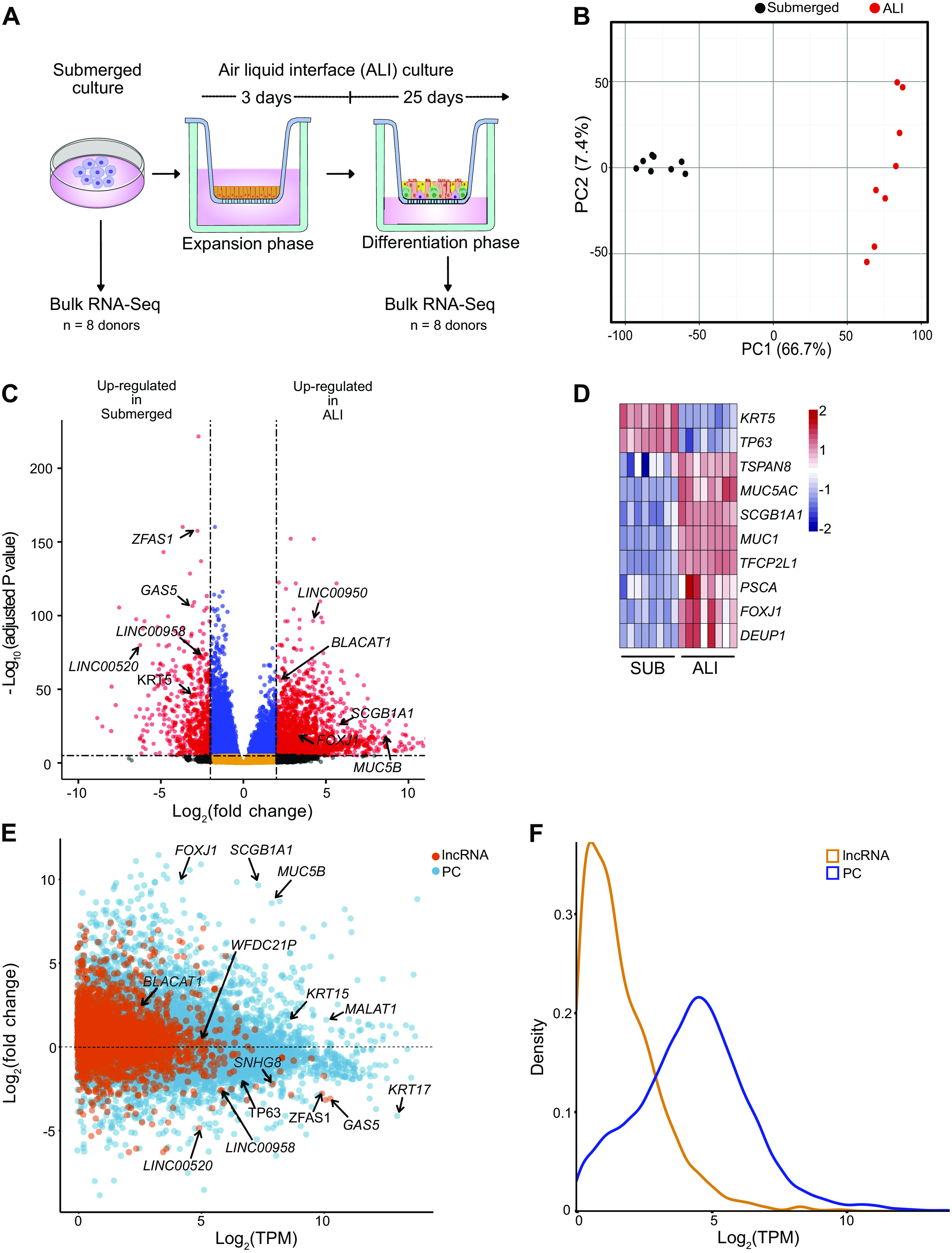
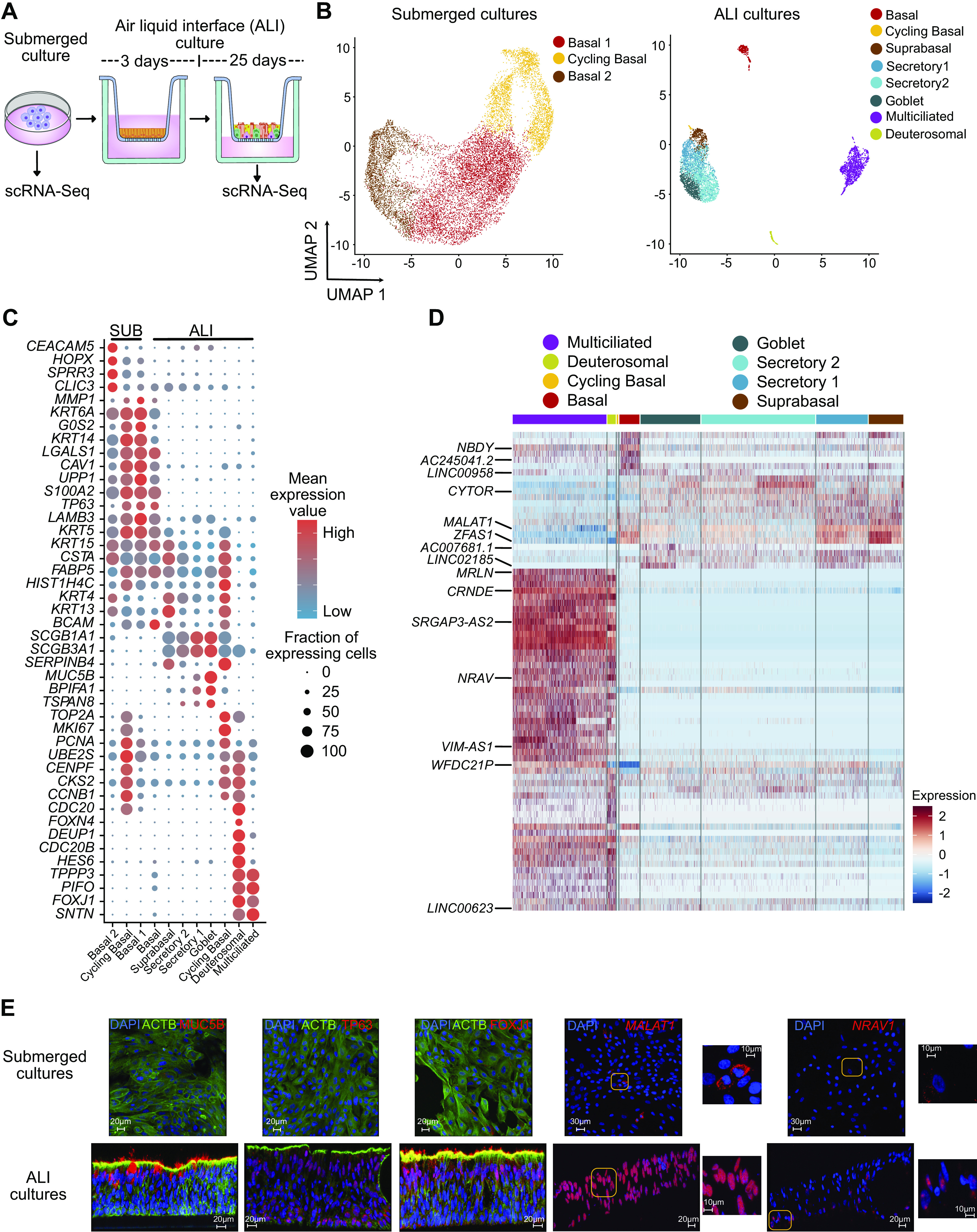
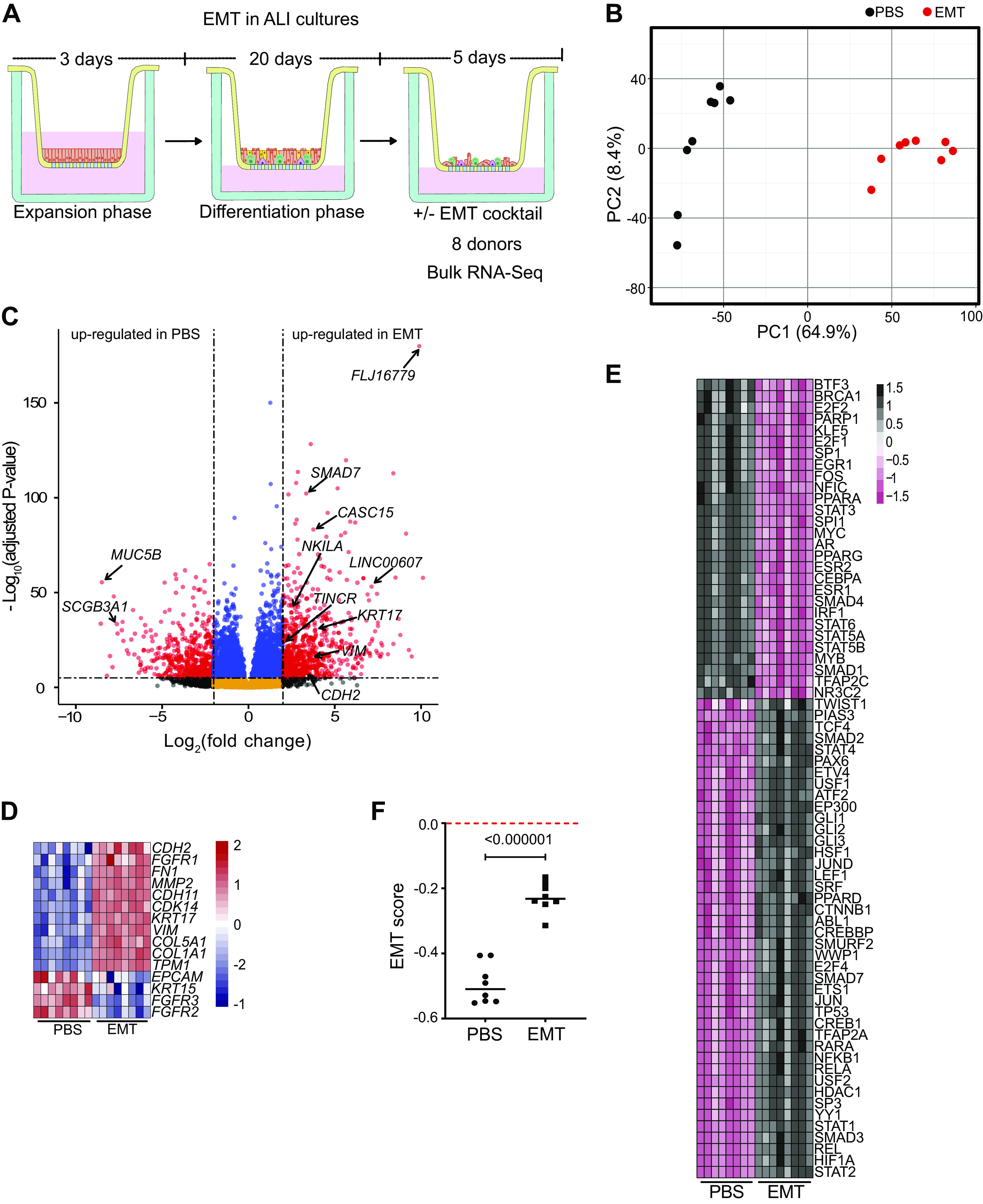
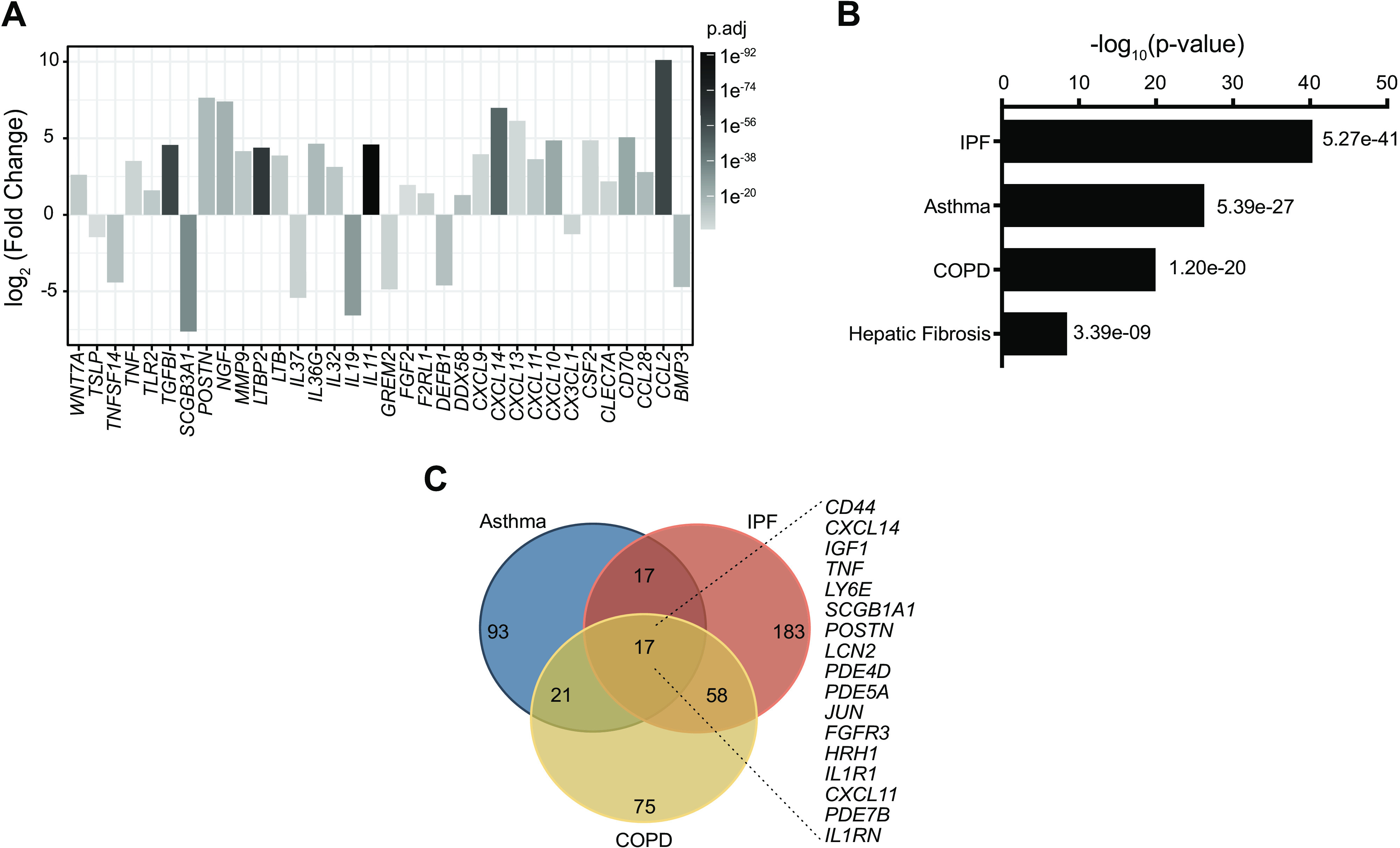
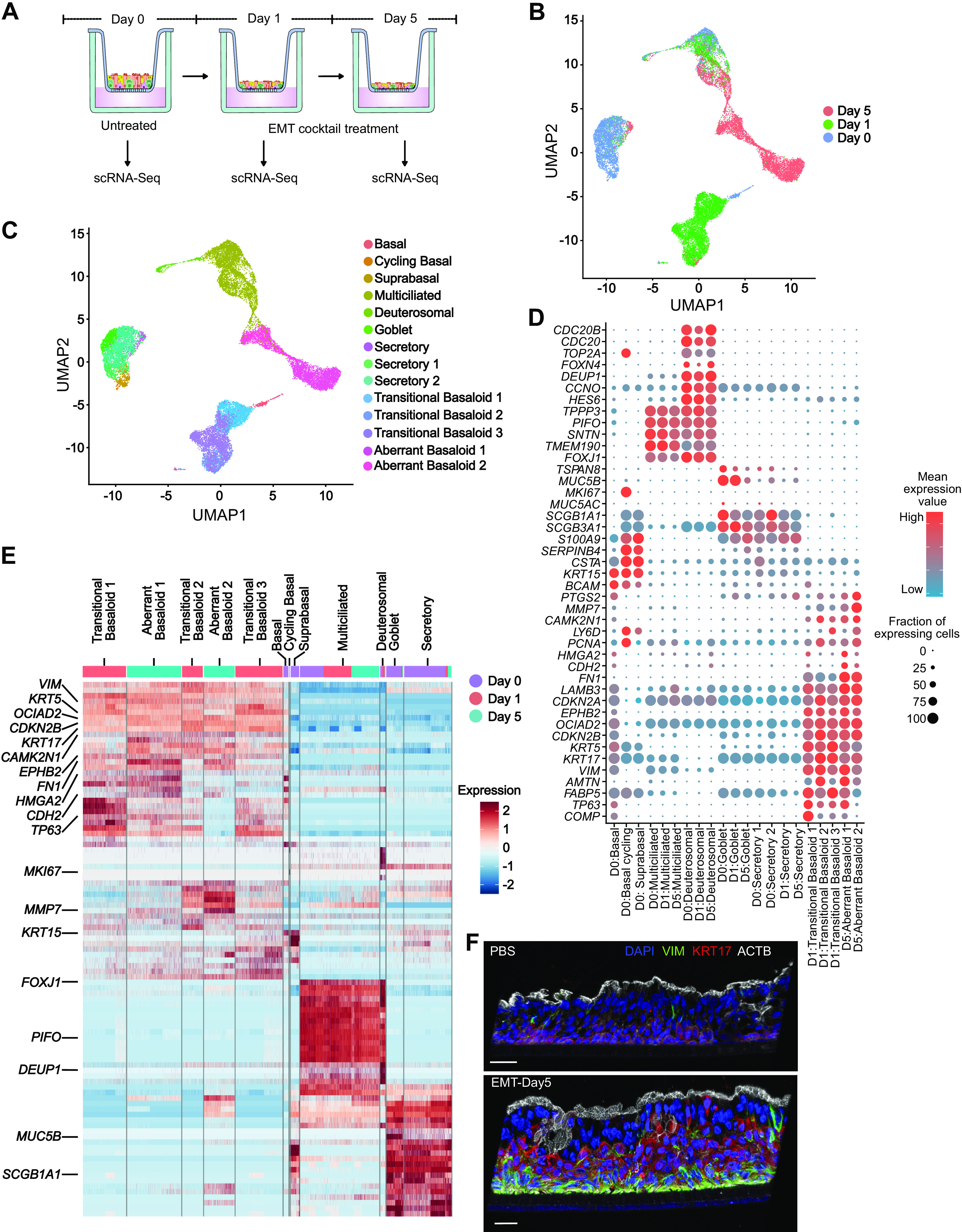
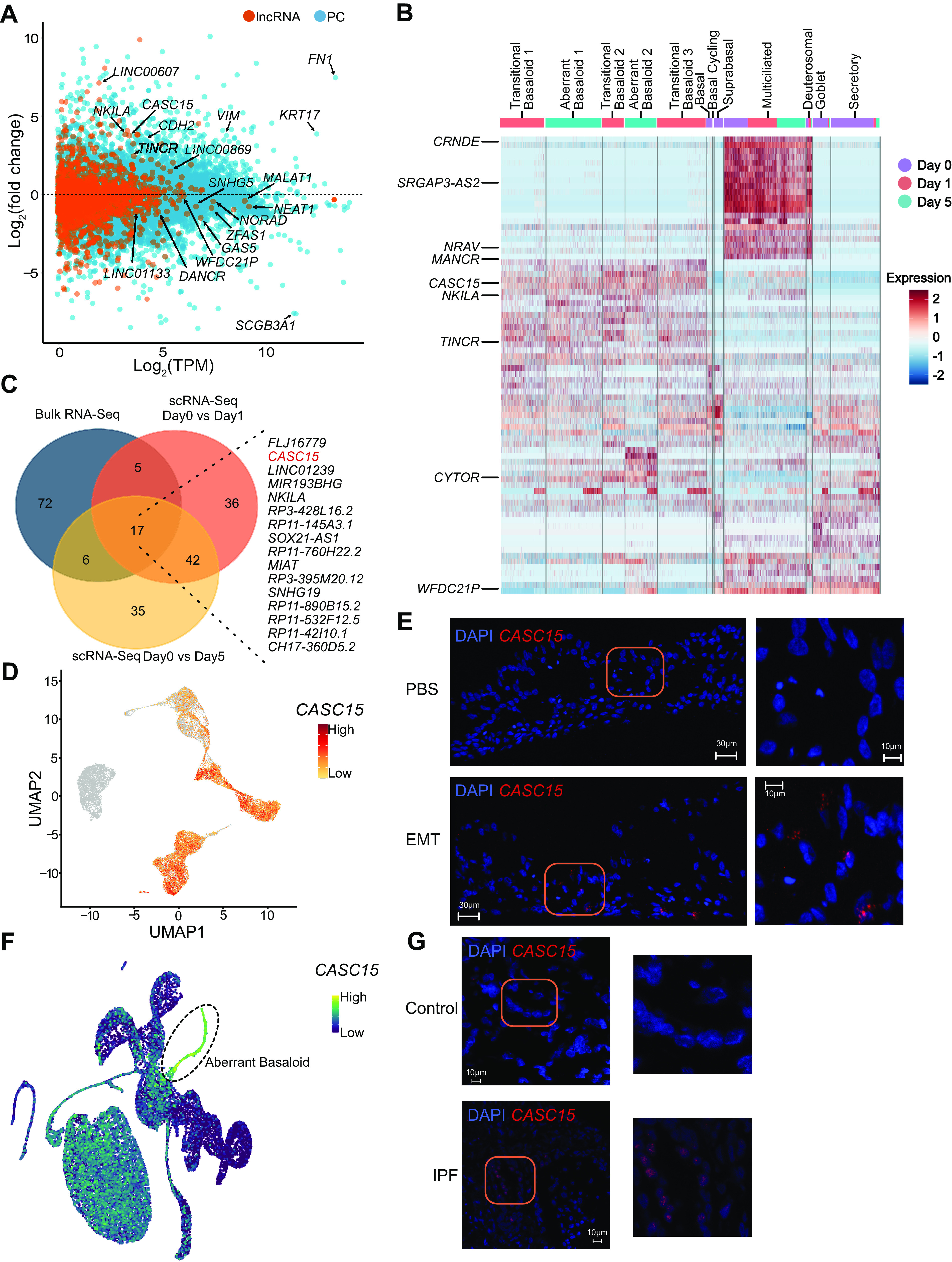
Although epithelial-mesenchymal transition (EMT) is a common feature of fibrotic lung disease, its role in fibrogenesis is controversial. Recently, aberrant basaloid cells were identified in fibrotic lung tissue as a novel epithelial cell type displaying a partial EMT phenotype. The developmental origin of these cells remains unknown. To elucidate the role of EMT in the development of aberrant basaloid cells from the bronchial epithelium, we mapped EMT-induced transcriptional changes at the population and single-cell levels. Human bronchial epithelial cells grown as submerged or air-liquid interface (ALI) cultures with or without EMT induction were analyzed by bulk and single-cell RNA-Sequencing. Comparison of submerged and ALI cultures revealed differential expression of 8,247 protein coding (PC) and 1,621 long noncoding RNA (lncRNA) genes and revealed epithelial cell-type-specific lncRNAs. Similarly, EMT induction in ALI cultures resulted in robust transcriptional reprogramming of 6,020 PC and 907 lncRNA genes. Although there was no evidence for fibroblast/myofibroblast conversion following EMT induction, cells displayed a partial EMT gene signature and an aberrant basaloid-like cell phenotype. The substantial transcriptional differences between submerged and ALI cultures highlight that care must be taken when interpreting data from submerged cultures. This work supports that lung epithelial EMT does not generate fibroblasts/myofibroblasts and confirms ALI cultures provide a physiologically relevant system to study aberrant basaloid-like cells and mechanisms of EMT. We provide a catalog of PC and lncRNA genes and an interactive browser (https://bronc-epi-in-vitro.cells.ucsc.edu/) of single-cell RNA-Seq data for further exploration of potential roles in the lung epithelium in health and lung disease.

Keywords: EMT; ILD; aberrant basaloid cells; epithelial-mesenchymal plasticity (EMP); fibrosis.


Conflict of interest statement

N. Kaminski served as a consultant to Boehringer Ingelheim, Third Rock, Pliant, Samumed, NuMedii, Theravance, LifeMax, Three Lake Partners, Optikira, Astra Zeneca, RohBar, Veracyte, Augmanity, CSL Behring, Galapagos, Gilead, and Thyron over the past 3 years, reports Equity in Pliant and Thyron, and received a grant from Veracyte, Boehringer Ingelheim, BMS, and nonfinancial support from MiRagen and Astra Zeneca. N. Kaminski has IP on novel biomarkers and therapeutics in IPF licensed to Biotech. None of the other authors has any conflicts of interest, financial or otherwise, to disclose.

Figures

Figure 1.
Figure 1.
Differentiation of the lung epithelium in vitro induces differential expression of thousands of genes including 1,621 lncRNAs. A: bulk RNA-Seq experimental design. HBECs from eight donors were cultured in submerged cultures and as ALI cultures before analysis by RNA-Seq. B: unbiased clustering of all samples using principal component analysis (PCA). All genes with <1 TPM value (averaged across all samples) were removed before performing PCA, n = 8 donors. C: volcano plot of expressed genes (TPM ≥ 1) between submerged and ALI cultures, n = 8 donors. Red dots, adjusted P value <10−6 and log2 fold-change >2; blue dots, adjusted P value <10−6 and log2 fold-change <2; black dots, adjusted P value >10−6 and log2 fold-change >2; orange dots, >10−6 and log2 fold-change <2. D: lung epithelial markers, TPM values plotted as heatmap between submerged (SUB) and ALI cultures, n = 8 donors. E: MA plot of protein coding (PC) and long noncoding RNA (lncRNA) expression between submerged and ALI cultures, n = 8. F: density plot depicting expression of PC and lncRNAs. Plotted are log2 TPM values averaged across submerged and ALI cultures from all donors, n = 8 donors. ALI, air-liquid interface; HBECs, human bronchial epithelial cells; PC, protein coding.
Figure 2.
Figure 2.
Single-cell analysis reveals epithelial cell-type-specific lncRNAs. A: schematic representation of submerged and ALI cultures. Primary human bronchial epithelial cells (HBECs) were dissociated from either submerged or ALI cultures before analysis by scRNA-Seq, n = 3 donors. B: UMAP plot of the scRNA-Seq expression data highlighting the main cell clusters observed in (left) submerged cultures and (right) ALI cultures. C: expression data highlighting selected cell-specific markers for cell clusters in submerged cultures and ALI cultures. D: heatmap depicting relative expression (normalized and scaled expression) of lncRNAs in each cluster in ALI cultures. All lncRNA names and their respective expression values are available in Supplemental Table S7. E: immunofluorescence (MUC5B, TP63, and FOXJ1) and RNA in situ hybridization (MALAT1 and NRAV) demonstrating expression of selected protein and RNA molecules in submerged and ALI cultures. Scales are depicted as micrometers; n = 2 donors, representative data from one donor is shown. ALI, air-liquid interface; lncRNA, long noncoding RNA; scRNA-Seq, single-cell RNA-sequencing.
Figure 3.
Figure 3.
Induction of EMT in ALI cultures induces a transcriptional reprogramming consistent with a partial EMT phenotype. A: bulk RNA-Seq experimental design. Differentiated ALI cultures treated with PBS or EMT cocktail for 5 days before RNA-Seq analysis. B: unbiased clustering of all samples using principal component analysis (PCA). All genes with <1 TPM value were removed before performing PCA, n = 8 donors. C: volcano plot of expressed genes (TPM ≥ 1) between PBS- and EMT-treated ALI cultures, n = 8. Red dots, adjusted P value <10−6 and log2 fold-change >2; blue dots, adjusted P value <10−6 and log2 fold-change <2; black dots, adjusted P value >10−6 and log2 fold-change >2; orange dots, >10−6 and log2 fold-change <2. D: EMT-associated markers, TPM values plotted as heatmap between PBS- and EMT-treated ALI cultures, n = 8 donors. E: NetAct analysis depicting activity of enriched transcription factors (FDR ≤ 0.01). Presence of TWIST1 and CTNNB1 but absence of major EMT markers like SNAI and ZEB family members indicate partial EMT. F: calculated EMT score for ALI cultures treated with PBS or EMT cocktail. A negative score for EMT-treated ALI cultures demonstrates a partial EMT. ALI, air-liquid interface; EMT, epithelial-mesenchymal transition; scRNA-Seq, single-cell RNA-sequencing.
Figure 4.
Figure 4.
Gene signatures from EMT-induced ALI cultures overlap those from different lung diseases. A: expression or immune associated genes in RNA-Seq data from ALI cultures treated with or without EMT cocktail, identified by Gene Ontology (GO) analysis. n = 8 donors. B: Ingenuity Pathway Analysis (IPA) of genes differentially expressed between untreated (control) and EMT-treated ALI cultures, compared with custom built disease gene lists. Plotted are P values for enrichment scores. C: Venn diagram for the genes identified from IPA analysis in asthma, IPF, and COPD. Listed are differentially expressed genes between untreated (control) and EMT-treated ALI cultures that also overlap all three diseases. ALI, air-liquid interface; COPD, chronic obstructive pulmonary disease; EMT, epithelial-mesenchymal transition.
Figure 5.
Figure 5.
Single-cell RNA-Seq of EMT in ALI cultures identifies aberrant basaloid-like cells but no fibroblast or myofibroblast conversion. A: scRNA-Seq experimental design. Differentiated HBEC ALI cultures (25 days) were dissociated for scRNA-Seq analysis following EMT treatment for 0, 1, and 5 days, n = 3 donors. B: UMAP plot of the scRNA-Seq expression data highlighting clusters defined by timepoints of EMT treatment. C: UMAP plot depicting cluster annotations of day 0, day 1, and day 5 EMT ALI cultures. D: expression data for selected genes for each cluster. Transitional basaloid-like and aberrant basaloid-like cells express both basal and mesenchymal markers. E: heatmap depicting relative expression (normalized and scaled z-scores) of PC genes in each cluster. All PC gene names, and their respective expression values is available in Supplemental Table S7. F: immunofluorescence demonstrating colocalization of VIM and KRT17 in EMT-treated ALI cultures. Scale = 30 µm; n = 2 donors, representative data from one donor is shown. ALI, air-liquid interface; EMT, epithelial-mesenchymal transition; HBECs, human bronchial epithelial cells; PC, protein coding.
Figure 6.
Figure 6.
EMT induction reprograms the lncRNA landscape. A: MA plot of protein coding (PC) and long noncoding RNA (lncRNA) expression between PBS- and EMT-treated ALI cultures, n = 8. B: heatmap depicting relative expression (normalized and scaled expression) of lncRNAs in each cluster. All lncRNA names and their respective expression values are available in Supplemental Table S7. Highlighted lncRNAs CASC15, TINCR, and MANCR exclusively mark transitional basaloid-like and aberrant basaloid-like cells. C: Venn diagram of top 100 lncRNA genes (as ranked based on adjusted P value) identified from bulk RNA-Seq, and pseudo-bulk analysis of scRNA-Seq of EMT-treated ALI cultures; listed are overlapping genes. D: UMAP plot highlighting expression of CASC15 in each cluster. E: in situ hybridization showing CASC15 expression in EMT-treated ALI cultures but not in PBS-treated ALI cultures. Scale = 30 µm; n = 2, representative data from one donor is shown. F: UMAP highlighting expression of CASC15 in IPF scRNA-Seq data. G: in situ hybridization using CASC15 probes on lung tissue from donors with or without IPF; scale = 30 µm; n = 1, data shown are from one donor each. IPF sample: 64 old, male, Caucasian. non-IPF (control) sample: 66 old, male, Caucasian. Confocal imaging demonstrates absence of CASC15 expression in non-IPF lung tissue but capturing punctuated signal in IPF lung tissue. ALI, air-liquid interface; EMT, epithelial-mesenchymal transition; lncRNA, long noncoding RNA.

References

    1. Lamouille S, Xu J, Derynck R. Molecular mechanisms of epithelial-mesenchymal transition. Nat Rev Mol Cell Biol 15: 178–196, 2014. doi:10.1038/nrm3758. - DOI - PMC - PubMed
    1. Dongre A, Weinberg RA. New insights into the mechanisms of epithelial-mesenchymal transition and implications for cancer. Nat Rev Mol Cell Biol 20: 69–84, 2019. doi:10.1038/s41580-018-0080-4. - DOI - PubMed
    1. Nieto MA. The ins and outs of the epithelial to mesenchymal transition in health and disease. Annu Rev Cell Dev Biol 27: 347–376, 2011. doi:10.1146/annurev-cellbio-092910-154036. - DOI - PubMed
    1. Sohal SS, Mahmood MQ, Walters EH. Clinical significance of epithelial mesenchymal transition (EMT) in chronic obstructive pulmonary disease (COPD): potential target for prevention of airway fibrosis and lung cancer. Clin Transl Med 3: 33, 2014. doi:10.1186/s40169-014-0033-2. - DOI - PMC - PubMed
    1. Nishioka M, Venkatesan N, Dessalle K, Mogas A, Kyoh S, Lin TY, Nair P, Baglole CJ, Eidelman DH, Ludwig MS, Hamid Q. Fibroblast-epithelial cell interactions drive epithelial-mesenchymal transition differently in cells from normal and COPD patients. Respir Res 16: 72, 2015. doi:10.1186/s12931-015-0232-4. - DOI - PMC - PubMed

Publication types

MeSH terms

Substances