. 2022 Apr-Jun;13(2):19476035221093060.
doi: 10.1177/19476035221093060.

Low-Intensity Pulsed Ultrasound Enhances the Efficacy of Bone Marrow-Derived MSCs in Osteoarthritis Cartilage Repair by Regulating Autophagy-Mediated Exosome Release

Affiliations

Low-Intensity Pulsed Ultrasound Enhances the Efficacy of Bone Marrow-Derived MSCs in Osteoarthritis Cartilage Repair by Regulating Autophagy-Mediated Exosome Release

Peng Xia et al. Cartilage. 2022 Apr-Jun.

Abstract

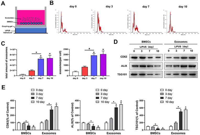
Objective: The present study explored whether low-intensity pulsed ultrasound (LIPUS) enhances the therapeutic efficacy of mesenchymal stem cells (MSCs) in osteoarthritis (OA) cartilage repair by regulating autophagy-mediated exosome release.

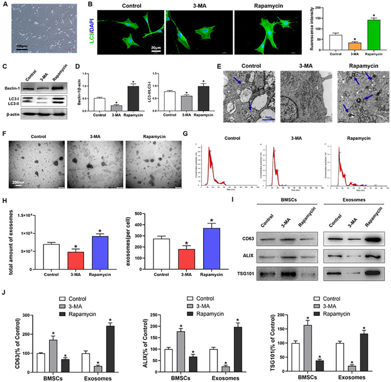
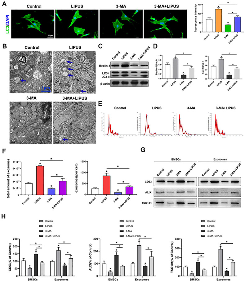
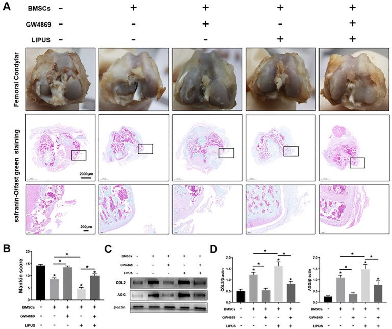
Design: MSCs were isolated from the rat bone marrow and treated with rapamycin, 3-methyladenine, or LIPUS. The mechanism of the LIPUS-stimulated exosome release by MSCs was analyzed by inhibiting autophagy. In addition, the MSCs were co-cultured with OA chondrocytes and stimulated by LIPUS, with or without exosome release inhibitor intervention. The exosome release was detected through transmission electron microscopy (TEM), nanoparticle tracking analysis, and biomarker expression analysis. Autophagy was analyzed through TEM, autophagy-related gene expression analysis, and immunofluorescence analysis in vitro. Furthermore, a rat knee OA model was constructed and treated with MSCs, GW4869, and LIPUS. The cartilage repair was assessed through histopathological analysis and extracellular matrix protein expression analysis.

Results: The in vitro results indicated that LIPUS promoted MSC exosome release by activating autophagy. The in vivo results demonstrated that LIPUS significantly enhanced the positive effects of MSCs on OA cartilage. These effects were significantly blocked by GW4869, an inhibitor of exosome release.

Conclusions: LIPUS can enhance the therapeutic efficacy of MSCs in OA cartilage repair, and the underlying mechanism is related to the increase in autophagy-mediated exosome release.

Keywords: autophagy; exosome; low-intensity pulsed ultrasound; mesenchymal stem cell; osteoarthritis.


Conflict of interest statement

Declaration of Conflicting Interests: The author(s) declared no potential conflicts of interest with respect to the research, authorship, and/or publication of this article.

Figures

Figure 1.
Figure 1.
Autophagy regulates exosome release by MSCs. (A) BMSCs cultured in medium; scale bars = 100 μm. (B) Immunofluorescence staining depicting LC3+ cells (green) and quantitative analysis of intensity; scale bars = 20 μm. (C, D) Western blot analysis of Beclin1, LC3I, LC3II, and β-actin expressions in BMSCs. (E) TEM depicting autophagosomes (arrows); scale bars = 1 μm. (F) TEM observation of exosomes from BMSCs; scale bars = 200 nm. (G, H) NTA of the size distribution and amount of exosomes from BMSCs. (I, J) Western blot analysis of CD63, ALIX and TSG101 expressions in BMSCs and exosomes. MSCs = mesenchymal stem cells; BMSCs = bone marrow-derived MSCs; TEM = transmission electron microscopy; NTA = nanoparticle tracking analysis; DAPI: diamidine phenylindole; 3-MA = 3-methyladenine. The values are the mean ± SD; n = 3, *P < .05.
Figure 2.
Figure 2.
LIPUS promotes MSC exosome release. (A) BMSCs were stimulated by LIPUS. (B, C) NTA of the size distribution and amount of exosomes from BMSCs. (D, E) Western blot analysis of CD63, ALIX, and TSG101 expressions in BMSCs and exosomes. LIPUS = low-intensity pulsed ultrasound; MSC = mesenchymal stem cell; BMSCs = Bone marrow-derived MSCs. The values are the mean ± SD; n = 3, * P < .05.
Figure 3.
Figure 3.
Inhibition of autophagy decreases the promoting effect of LIPUS on MSC exosome release. (A) Immunofluorescence staining depicting LC3+ cells (green) and quantitative analysis of intensity; scale bars = 20 μm. (B) Electron microscopy depicting autophagosomes (arrows); scale bars = 1 μm. (C, D) Western blot analysis of Beclin1, LC3I, LC3II, and β-actin expressions in BMSCs. (E, F) NTA of the size distribution and amount of exosomes from BMSCs. (G, H) Western blotting analysis of CD63, ALIX, and TSG101 expressions in BMSCs and exosomes. LIPUS = low-intensity pulsed ultrasound; MSC = mesenchymal stem cell; BMSCs = bone marrow-derived MSCs; NTA = nanoparticle tracking analysis; DAPI: diamidine phenylindole; 3-MA = 3-methyladenine. The values are the mean ± SD; n = 3, *P < .05.
Figure 4.
Figure 4.
LIPUS enhances the anti-degeneration effects of MSCs on OA chondrocytes by promoting exosome release. (A) The OA chondrocyte and BMSC co-culture system was stimulated by LIPUS. (B) Immunohistochemical staining (ICC) and toluidine blue staining in OA chondrocytes. (C, D) Western blotting analysis of COL2, AGG, and β-actin expressions in OA chondrocytes. LIPUS = low-intensity pulsed ultrasound; MSCs = mesenchymal stem cells; OA = osteoarthritis; BMSC = bone marrow-derived MSC; COL2 = type II collagen; AGG = aggrecan. The values are the mean ± SD; n = 3, *P < .05.
Figure 5.
Figure 5.
LIPUS enhances the repair effects of MSCs on OA cartilage through the exosome release pathway. (A) The femoral condylar cartilage was observed and detected through safranin-O/fast green staining; scale bars = 2,000 μm, 200 μm. (B) Bar graph comparing the Mankin scores. (C, D) Western blotting analysis of COL2, AGG, and β-actin expressions in the cartilage. LIPUS = low-intensity pulsed ultrasound; MSCs = mesenchymal stem cells; OA = osteoarthritis; COL2 = type II collagen; AGG = aggrecan. The values are the mean ± SD; n = 6, *P < .05.

Similar articles

Cited by

References

    1. Bortoluzzi A, Furini F, Scirè CA. Osteoarthritis and its management—epidemiology, nutritional aspects and environmental factors. Autoimmun Rev. 2018;17(11):1097-104. doi:10.1016/j.autrev.2018.06.002. - DOI - PubMed
    1. Hunter DJ, Bierma-Zeinstra S. Osteoarthritis. Lancet. 2019;393(10182):1745-59. doi:10.1016/S0140-6736(19)30417-9. - DOI - PubMed
    1. Nelson AE, Allen KD, Golightly YM, Goode AP, Jordan JM. A systematic review of recommendations and guidelines for the management of osteoarthritis: the chronic osteoarthritis management initiative of the U.S. bone and joint initiative. Semin Arthritis Rheum. 2014;43(6):701-12. doi:10.1016/j.semarthrit.2013.11.012. - DOI - PubMed
    1. Block JA. Osteoarthritis: OA guidelines: improving care or merely codifying practice? Nat Rev Rheumatol. 2014;10(6):324-6. doi:10.1038/nrrheum.2014.61. - DOI - PubMed
    1. Chen FH, Rousche KT, Tuan RS. Technology insight: adult stem cells in cartilage regeneration and tissue engineering. Nat Clin Pract Rheumatol. 2006;2(7):373-82. doi:10.1038/ncprheum0216. - DOI - PubMed

Publication types