. 2022 Aug;19(4):809-821.
doi: 10.1007/s13770-022-00443-7. Epub 2022 Apr 19.

Asymmetric Contribution of Blastomere Lineages of First Division of the Zygote to Entire Human Body Using Post-Zygotic Variants

Affiliations

Asymmetric Contribution of Blastomere Lineages of First Division of the Zygote to Entire Human Body Using Post-Zygotic Variants

Seong Gyu Kwon et al. Tissue Eng Regen Med. 2022 Aug.

Abstract

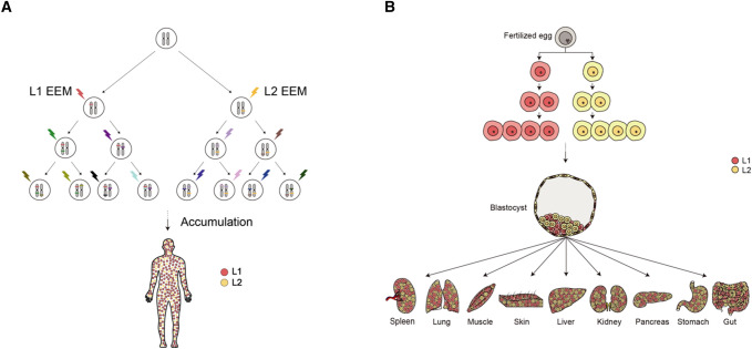
Background: In humans, after fertilization, the zygote divides into two 2n diploid daughter blastomeres. During this division, DNA is replicated, and the remaining mutually exclusive genetic mutations in the genome of each cell are called post-zygotic variants. Using these somatic mutations, developmental lineages can be reconstructed. How these two blastomeres are contributing to the entire body is not yet identified. This study aims to evaluate the cellular contribution of two blastomeres of 2-cell embryos to the entire body in humans using post-zygotic variants based on whole genome sequencing.

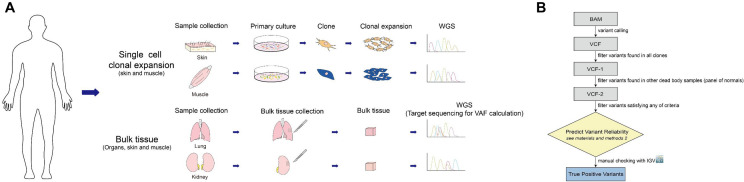
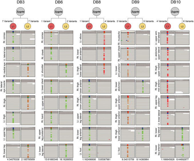
Methods: Tissues from different anatomical areas were obtained from five donated cadavers for use in single-cell clonal expansion and bulk target sequencing. After conducting whole genome sequencing, computational analysis was applied to find the early embryonic mutations of each clone. We developed our in-house bioinformatics pipeline, and filtered variants using strict criteria, composed of mapping quality, base quality scores, depth, soft-clipped reads, and manual inspection, resulting in the construction of embryological phylogenetic cellular trees.

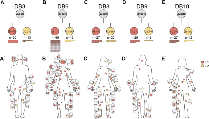
Results: Using our in-house pipeline for variant filtering, we could extract accurate true positive variants, and construct the embryological phylogenetic trees for each cadaver. We found that two daughter blastomeres, L1 and L2 (lineage 1 and 2, respectively), derived from the zygote, distribute unequally to the whole body at the clonal level. From bulk target sequencing data, we validated asymmetric contribution by means of the variant allele frequency of L1 and L2. The asymmetric contribution of L1 and L2 varied from person to person.

Conclusion: We confirmed that there is asymmetric contribution of two daughter blastomeres from the first division of the zygote across the whole human body.

Keywords: Asymmetry; Clonal expansion; Early embryonic mutations; Lineage tracing; Mutation filtering; Somatic mutation.


Conflict of interest statement

The authors declares that they have no conflict of interest.

Figures

Fig. 1
Fig. 1
Overview of the process. A Experimental design. The main purpose for sample collection was for single-cell clonal expansion, and bulk tissue target sequencing. WGS was conducted on every single-cell clonal expansion sample. B Computational analysis pipeline. Detailed workflow consists of two main parts. First, mutations that are present in same or other DB samples were filtered using information in variant call format (VCF) files only. Second, mutations that met specific criteria were filtered using information in BAM files
Fig. 2
Fig. 2
Lineage tree for each dead body in 1st cell division. IGV shows reads, depths, variant information visually using BAM files as input. Vertical lines on the upper side of the IGV snapshots represent depth of the reads in the position of the variants. Variants are presented as colored points. Green: alt = A, red: alt = T, brown: alt = G, blue: alt = C. X-axis represents genomic locations of the variants (Chrom:Pos) and y-axis represents the name of samples at different anatomical locations. For example, in DB3, variant 4:34,076,338 is present in clones of left lateral eye, left upper leg, left thigh, right lateral eye muscle, and right cornea, but not in  clones of right lower leg, right medial eye, and the second right lower leg
Fig. 3
Fig. 3
A-E Clonal diversity of L1 and L2 in whole body.A'-E' Distribution of cells of L1 and L2 are presented in red and yellow dots. For A–E, percentages, and number of clones of L1 and L2 are presented. For A’–E’, proportion of clones in each location is presented in pie charts and detailed numbers are in magnified ovals
Fig. 4
Fig. 4
VAF distribution in the various tissues in each DB with pie charts. Pie charts are located in each anatomical tissue in DBs and show VAFs in terms of L1 and L2. Mean VAFs for skin, muscle, fat, organs, and others are presented in bar charts
Fig. 5
Fig. 5
Schematic overview. A Somatic mutations are accumulated for each division. All of the accumulated mutations remain in the cells as the division proceeds. Therefore, all of the cells in the whole body can be classified into L1 or L2 related cells [18]. B The development of a human began from a fertilized egg. As a zygote divides into two blastomeres, those cells will be the ancestors for all organs in the human body. First, cells from L1 and L2 divides into different early lineages. Cells are then mixed to form a blastocyst, which consists of ICM and the trophoblast. ICM is the precursor for all the cells in the body, and certain populations of the cells are lost in the trophoblast placenta. Eventually, all of the organs consist of the mixed asymmetric populations of L1 and L2 originated cells in variable ratios

References

    1. Gerri C, Menchero S, Mahadevaiah SK, Turner JMA, Niakan KK. Human embryogenesis: a comparative perspective. Annu Rev Cell Dev Biol. 2020;36(1):411–440. doi: 10.1146/annurev-cellbio-022020-024900. - DOI - PubMed
    1. Sender R, Fuchs S, Milo R. Revised estimates for the number of human and bacteria cells in the body. PLoS Biol. 2016;14(8):e1002533. doi: 10.1371/journal.pbio.1002533. - DOI - PMC - PubMed
    1. Shahbazi MN, Zernicka-Goetz M. Deconstructing and reconstructing the mouse and human early embryo. Nat Cell Biol. 2018;20(8):878–887. doi: 10.1038/s41556-018-0144-x. - DOI - PubMed
    1. Wang H, Dey SK. Roadmap to embryo implantation: clues from mouse models. Nat Rev Genet. 2006;7:185–99. - PubMed
    1. Rossant J. Mouse and human blastocyst-derived stem cells: vive les differences. Development. 2015;142:9–12. - PubMed

Publication types

LinkOut - more resources