Review
. 2022 Dec;25(4):783-797.
doi: 10.1007/s40477-021-00619-2. Epub 2022 Apr 19.

Non-glandular findings on breast ultrasound. Part I: a pictorial review of superficial lesions

Affiliations
Review

Non-glandular findings on breast ultrasound. Part I: a pictorial review of superficial lesions

Martina Caruso et al. J Ultrasound. 2022 Dec.

Abstract

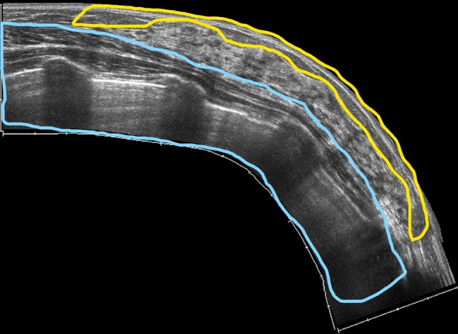
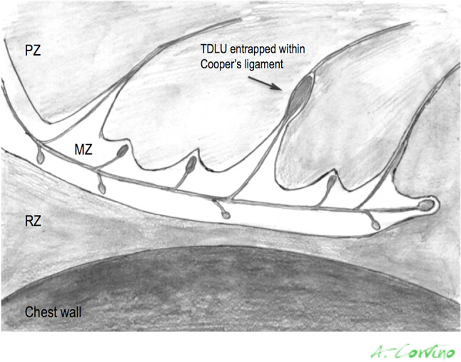
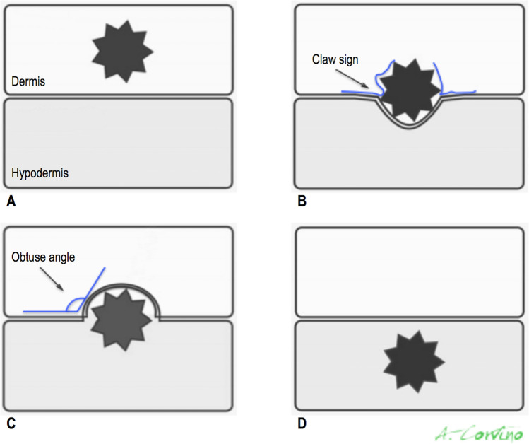
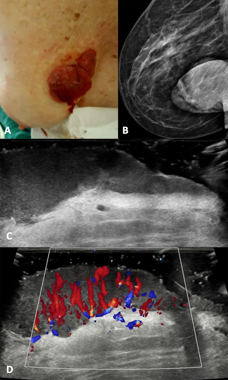
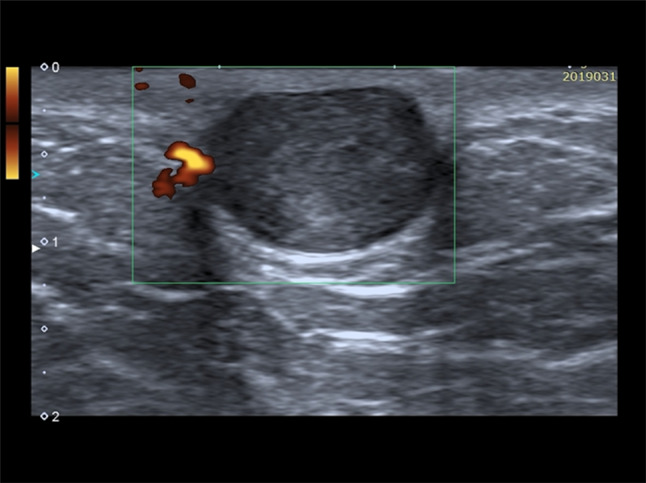
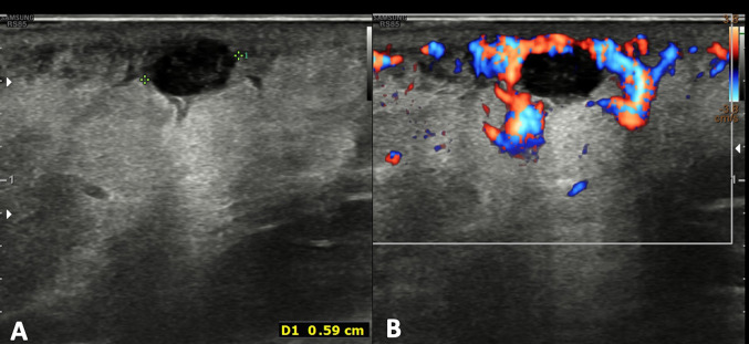
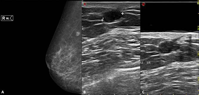
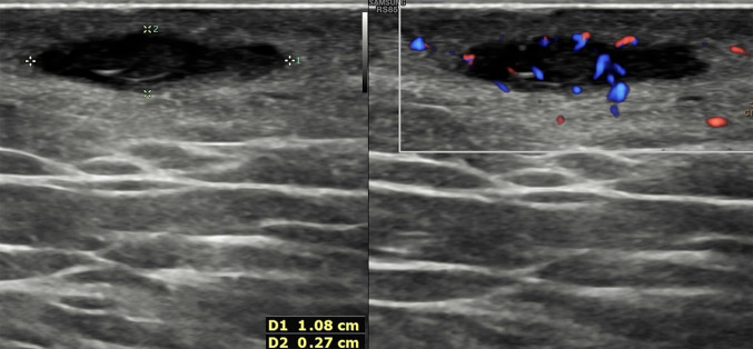
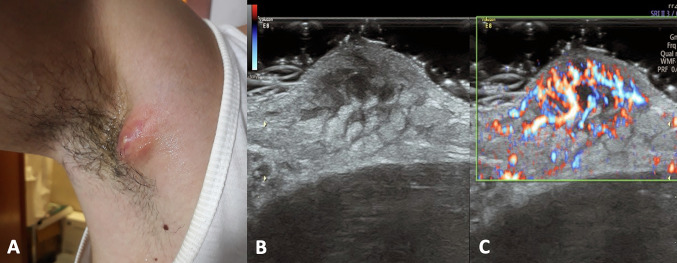
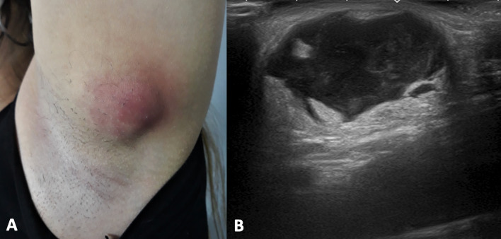
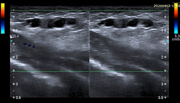
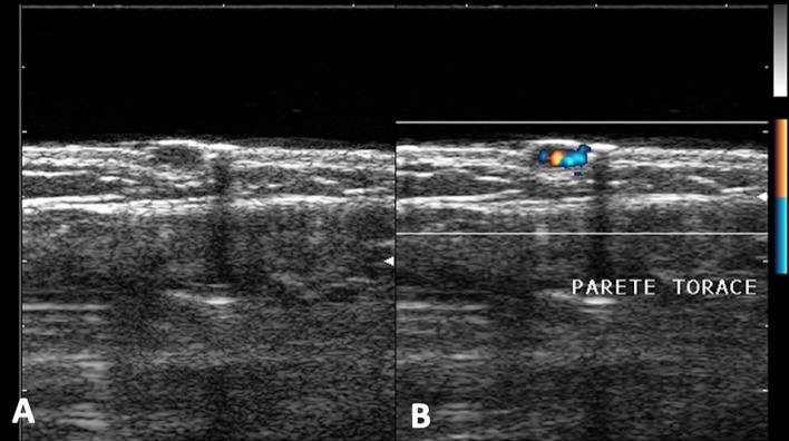
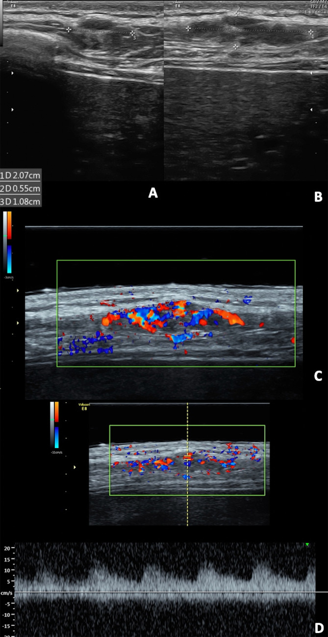
Ultrasound (US) represents the first-level imaging technique in the assessment of breast in young patients, whereas it is complementary to mammography in adult ones. It is not uncommon to encounter non-glandular mass during either screening or diagnostic breast US; sometimes the evaluation of palpable lump may be the reason of clinician's US request. The breast US field-of-view includes not only the glandular parenchyma, but also the tissues located anterior and posterior to it, from the skin to the ribs. In this setting, the radiologist must be familiar with the non-glandular breast diseases, which can occur in the superficial layers as well as in the chest wall. The differential diagnosis varies according to anatomic layer, so the anatomic origin is the first feature to assess and the correct localization is needed to avoid misdiagnosis and to choose, when requested, the second diagnostic step, imaging or histologic analysis. This paper is the first of two focused on non-glandular breast lesions; characterization, differential diagnosis, and pitfalls of superficial lesions are reviewed. They may be located in the dermis or hypodermis: the former are usually benign skin lesions, whereas the latter, although usually benign, may arise also from the anterior terminal lobular units, hence the papilloma, fibroadenomas, and breast cancers should be included in the differential diagnosis. US is more sensitive than CT and MRI in the assessment of superficial lesions due to higher spatial resolution.

Keywords: Breast Ultrasound; Characterization; Differential Diagnosis; Hypodermal Lesions; Pitfalls; Skin Lesions; Superficial non-glandular Lesions.


Conflict of interest statement

We confirm that this work is original and has not been published elsewhere nor is it currently under consideration for publication elsewhere. Publication is approved by all authors and by the responsible authorities where the work was carried out. Each author have participated sufficiently in any submission to take public responsibility for its content. The authors have no conflicts of interest. Written informed consent was obtained from all patients, and the study was approved by the ethics committee of the institution.

Figures

Fig. 1
Fig. 1
US appearance of the breast region: pre-mammary zone (yellow box), mammary zone (between yellow and light blue boxes), and retro-mammary zones (light blue box)
Fig. 2
Fig. 2
Schematic picture of the breast region: pre-mammary zone (PZ), mammary zone (MZ), and retro-mammary zones (RZ). The superficial extensions of Cooper ligaments, structural support of the breast, cross the hypodermis and they may entrap some TDLUs
Fig. 3
Fig. 3
Diagram illustrates how to determine whether a lesion is dermal (A and B) or hypodermal (C and D) on the basis of US location and findings. Claw sign is determined by a “claw” of hypoechoic skin invaginating around the margins and supports dermal origin (B). Hypodermal lesions form obtuse angles with dermal line (C)
Fig. 4
Fig. 4
Clinical presentation, mammography (B), B-mode (C) and color Doppler (D) scans with a gel spacer. (A) Squamous cell carcinoma presents as red and raised nodule with some bleeding areas. (B) The mammography shows a large radiopaque nodule. (B, C) At US, it appears as an irregular skin thickness tending to involve the deeper layer with diffuse increased vascular signals.
Fig. 5
Fig. 5
Power Doppler scan of an epidermoid cyst. Oval, well-defined epidermoid cyst with slightly hypoechoic echotexture compared to dermal layer. Power Doppler does not show intralesional vascular signal. The “claw” sign confirms the dermal origin of the lesion
Fig. 6
Fig. 6
B-mode (A) and color Doppler (B) scans of the breast region. Oval epidermoid cyst with slightly irregular margins and hypo-anechoic internal echotexture. The surrounding dermal layer is thickened and hyperechoic due to edema, with increased vascular signal at color Doppler assessment
Fig. 7
Fig. 7
Right medio-lateral mammography (A), B-mode scans of the right breast without (B) and with (C) a gel stand-off pad. Oval, well-defined epidermoid cyst, which appears as an opacity at mammography, while a hypoechoic nodule at US, located in the hypodermal layer. The presence of the “punctum” (C) confirm the diagnosis of epidermoid cyst
Fig. 8
Fig. 8
Split screen US image showing B-mode scan of the axillary on the left and corrisponding color Doppler one on the right. A dermal fluid collection with echogenic fragments of hair is typical of hidradenitis suppurative. The surrounding tissues show an increased echogenicity and vascular signals at color Doppler due to inflammation and invasive behavior of the disease
Fig. 9
Fig. 9
Color Doppler scan of axillary in a child. Pilomatrixoma appears as an ovoid, hyper-vascular lesion, parallel to the skin and located at the dermis-hypodermis junction; the internal echotexture is heterogeneous, predominantly hypoechoic
Fig. 10
Fig. 10
Clinical presentation (A) and B-mode scan (B) of breast skin hematoma. The US appearance is represented by hyperechoic collection located in the hypodermis with some anechoic areas
Fig. 11
Fig. 11
Clinical presentation (A) and color Doppler scan (B) of an abscess due to ingrown hair. Physical examination shows a red bump and the skin around feel painful and warm to touch. US shows an anechoic fluid collection located in the hypodermis with a thin hyperechoic stria inside (hair ingrown). The surrounding tissues are thickened and inflamed with increased vascular signal at color Doppler
Fig. 12
Fig. 12
B-mode scans of the breast. A piercing fragment retained in the subcutaneous tissues appears hyperechoic with reverberation artifact and postero-lateral acoustic shadowings
Fig. 13
Fig. 13
Clinical presentation (A), B-mode (B) and color Doppler scans (C) of right axillary. Physical examination shows a slight red, painful bump. At US, the subcutaneous abscess appears as an anechoic area with indistinct margins without vascular signal at color Doppler; the surrounding tissues are very thickened, heterogenous, slightly hyperechoic due to edema (cobblestone appearance) and hypervascularized
Fig. 14
Fig. 14
Clinical presentation (A) and B-mode scan (B) of left axillary. Physical examination shows a red, painful bump, the surrounding area is swelled and warm to touch. Subcutaneous abscess presents as a fluid collection with irregular margins and heterogeneous internal echotexture because of the presence of debris. The surrounding tissue are slightly hyperechoic due to inflammation
Fig. 15
Fig. 15
Color Doppler scans of the breast. US appearance of Mondor’s disease is characterized by hypoechoic subcutaneous and uncompressible “rosary-like” structure with some internal hyperechoic area due to blood clots. No vascular flow is visible with Color Doppler
Fig. 16
Fig. 16
B-mode (A) and color Doppler (B) of the pectoral region in a child. Well-defined, hypoechoic cutaneous angioma with rich vascularization at color Doppler
Fig. 17
Fig. 17
B-mode (A and B), color Doppler (C) and spectral Doppler (D) scans. Lobulated, well-circumscribed and predominantly hypoechoic hemangioma with rich intralesional vascularization on color Doppler and low resistance arterial flow on spectral Doppler

References

    1. Giess CS, Raza S, Birdwell RL. Distinguishing breast skin lesions from superficial breast parenchymal lesions: Diagnostic criteria, imaging characteristics, and pitfalls. Radiographics. 2011;31:1959–1972. doi: 10.1148/rg.317115116. - DOI - PubMed
    1. Draghi F, Cocco G, Richelmi FM, Schiavone C. Abdominal wall sonography: a pictorial review. J Ultrasound. 2020;23:265–278. doi: 10.1007/s40477-020-00435-0. - DOI - PMC - PubMed
    1. Ji HY, Kim EK, Min JK, Ki KO. Imaging findings of chest wall lesions on breast sonography. J Ultrasound Med. 2008;27:125–138. doi: 10.7863/jum.2008.27.1.125. - DOI - PubMed
    1. Pandya S, Moore RG. Breast development and anatomy. Clin Obstet Gynecol. 2011;54:91–95. doi: 10.1097/GRF.0b013e318207ffe9. - DOI - PubMed
    1. Corvino A, Sandomenico F, Corvino F, et al. Utility of a gel stand-off pad in the detection of Doppler signal on focal nodular lesions of the skin. J Ultrasound. 2020;23:45–53. doi: 10.1007/s40477-019-00376-3. - DOI - PMC - PubMed