. 2022 Sep;74(9):1544-1555.
doi: 10.1002/art.42146. Epub 2022 Aug 22.

Interleukin-13 Receptor α1-Mediated Signaling Regulates Age-Associated/Autoimmune B Cell Expansion and Lupus Pathogenesis

Affiliations

Interleukin-13 Receptor α1-Mediated Signaling Regulates Age-Associated/Autoimmune B Cell Expansion and Lupus Pathogenesis

Zhu Chen et al. Arthritis Rheumatol. 2022 Sep.

Abstract

Objective: Age-associated/autoimmune B cells (ABCs) are an emerging B cell subset with aberrant expansion in systemic lupus erythematosus. ABC generation and differentiation exhibit marked sexual dimorphism, and Toll-like receptor 7 (TLR-7) engagement is a key contributor to these sex differences. ABC generation is also controlled by interleukin-21 (IL-21) and its interplay with interferon-γ and IL-4. This study was undertaken to investigate whether IL-13 receptor α1 (IL-13Rα1), an X-linked receptor that transmits IL-4/IL-13 signals, regulates ABCs and lupus pathogenesis.

Methods: Mice lacking DEF-6 and switch-associated protein 70 (double-knockout [DKO]), which preferentially develop lupus in females, were crossed with IL-13Rα1-knockout mice. IL-13Rα1-knockout male mice were also crossed with Y chromosome autoimmune accelerator (Yaa) DKO mice, which overexpress TLR-7 and develop severe disease. ABCs were assessed using flow cytometry and RNA-Seq. Lupus pathogenesis was evaluated using serologic and histologic analyses.

Results: ABCs expressed higher levels of IL-13Rα1 than follicular B cells. The absence of IL-13Rα1 in either DKO female mice or Yaa DKO male mice decreased the accumulation of ABCs, the differentiation of ABCs into plasmablasts, and autoantibody production. Lack of IL-13Rα1 also prolonged survival and delayed the development of tissue inflammation. IL-13Rα1 deficiency diminished in vitro generation of ABCs, an effect that, surprisingly, could be observed in response to IL-21 alone. RNA-Seq revealed that ABCs lacking IL-13Rα1 down-regulated some histologic characteristics of B cells but up-regulated myeloid markers and proinflammatory mediators.

Conclusion: Our findings indicate a novel role for IL-13Rα1 in controlling ABC generation and differentiation, suggesting that IL-13Rα1 contributes to these effects by regulating a subset of IL-21-mediated signaling events. These results also suggest that X-linked genes besides TLR7 participate in the regulation of ABCs in lupus.


Figures

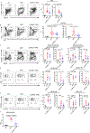
Figure 1.
Figure 1.. Lack of IL-13Rα1 decreases ABC accumulation and autoantibody production in DKO females.
Representative FACS plots and quantifications of a, CD11c+Tbet+ ABCs (gated on B220+) from the spleens of aged (24 wk) WT, DKO, and Il13rα1−/−DKO female mice. b, total GC B cells (gated on B220+ GL7+Fas+ splenocytes). c, total PB/PC (B220mid/loCD138+). d, CD11c+GL7+Fas+ cells (gated on B220+ splenocytes). e, CD11c+ PB/PC (B220mid/loCD138+). f, anti-dsDNA IgG2c levels in the serum as assessed by ELISA. Data show 7–20 mice that represent the total number of mice per group pooled from seven independent experiments (sample size derived from previous studies [21]) (a, b, c, e); 5–16 mice that represent the total number of mice per group pooled from five independent experiments (d); 9–12 mice that represent the total number of mice per group pooled from three independent experiments (f). All data show mean +/− SEM. *P<0.05, **P<0.01, ***P<0.001, ****P<0.0001 (one-way ANOVA followed by Bonferroni’s multiple-comparisons test).
Figure 2.
Figure 2.. IL-13Rα1 deficiency diminishes TLR7-driven ABC expansion and humoral responses in Yaa-DKO male mice.
a, Representative FACS plots and quantifications of splenic CD11c+Tbet+ ABCs (gated on B220+) of WT, YaaDKO and YaaIl13rα1−/YDKO males (>20 wk). b, Representative FACS plots and quantifications of total GC B cells (gated on B220+ GL7+Fas+ splenocytes) from WT, YaaDKO and YaaIl13rα1−/YDKO males (>20 wk). c, Representative FACS plots and quantifications of CD11c+GL7+Fas+ cells (gated on B220+ splenocytes). d, Representative FACS plots and quantifications of total splenic PB/PC (B220mid/loCD138+). e, Representative FACS plots and quantifications of CD11c+ PB/PC (B220mid/loCD138+). f, Anti-dsDNA IgG2c, anti-dsDNA IgG1, anti-SmRNP, anticardiolipin IgG, and anti-phosphatidylserine (PS) IgG serum levels assessed by ELISA. Data show 7–12 mice that represent the total number of mice per group pooled from seven independent experiments (a, b, d); 5–11 mice that represent the total number of mice per group pooled from five independent experiments (c); 5–12 mice that represent the total number of mice per group pooled from five independent experiments (e); 7–30 mice that represent the total number of mice per group pooled from three independent experiments (f.) All data show mean +/− SEM; *P<0.05, **P<0.01, ***P<0.001, ****P<0.0001 (one-way ANOVA followed by Bonferroni’s multiple-comparisons test).
Figure 3.
Figure 3.. IL-13Rα1-mediated signaling regulates ABC formation in vitro.
a, Representative FACS plot of CD11c and T-bet expression by CD23+ B cells purified from WT, DKO and Il13rα1−/−DKO female mice (6–10 weeks) stimulated for 3 d with anti-IgM (5μg/ml) and anti-CD40 (5μg/ml) alone or together with IL-21 (50ng/ml), IL-4 (10ng/ml), and IL-13 (20ng/ml). Numbers in quadrants indicate percent cells in each. b, Quantification of CD11c+T-bet+ ABCs in the cultures described in a. c, Quantification of CD11c expression in CD23+ B cells stimulated with anti-IgM, anti-CD40, and IL-21 described in a. Data show 6–8 mice that represent the total number of mice per group pooled from three independent experiments (a-c). **P<0.01, ***P<0.001, **** P<0.0001 (one-way ANOVA followed by Bonferroni’s multiple-comparisons test). d, Western blot of phosphorylated Stat6 (pStat6) and phosphorylated Stat3 (pStat3) from nuclear extracts of cells stimulated with anti-IgM (5μg/ml) and anti-CD40 (5μg/ml) alone or together with IL-21 (50ng/ml), IL-4 (10ng/ml), and IL-13 (20ng/ml) as described in a. Reprobing with HDAC1 was used as a loading control. Data are representative of two independent experiments.
Figure 4.
Figure 4.. DKO ABCs lacking IL-13Rα1 exhibit enhanced myeloid and proinflammatory features.
a-g, RNA-seq analysis was performed on RNA from ABCs (B220+CD19+CD11c+CD11b+) sorted by flow cytometry from DKO (n=5) and Il13rα1−/−DKO female mice (n=3) (>24 wk). a, Volcano plot showing differentially expressed genes (change in expression of over one-fold) in cells from DKO female mice relative to their expression in cells from Il13rα1−/−DKO female mice, plotted against FDR-corrected P value (P < 0.05; dashed horizontal line indicates cutoff of P = 0.05). b-c, Plot showing the top pathways upregulated (b) or downregulated (c) in Il13rα1−/−DKO ABCs by IPA. Dotted line indicates significance threshold at FDR q <0.25. Data were presented as z scores of expression by k means clustering of log transformed expression (counts per million) of genes expressed differentially by these cells.d-e, Top upstream transcription factors upregulated (d) or downregulated (e) in Il13rα1−/−DKO ABCs as predicted by IPA. f, Heatmap shoring differentially expressed genes in Hallmark Inflammatory Response pathway. g, Heatmap showing differentially expressed genes in Hallmark Epithelial Mesenchymal Transition.

References

    1. Liu Z and Davidson A, Taming lupus-a new understanding of pathogenesis is leading to clinical advances. Nat Med, 2012. 18(6): p. 871–82. - PMC - PubMed
    1. Tsokos GC, Autoimmunity and organ damage in systemic lupus erythematosus. Nat Immunol, 2020. 21(6): p. 605–614. - PMC - PubMed
    1. Seth A and Craft J, Spatial and functional heterogeneity of follicular helper T cells in autoimmunity. Curr Opin Immunol, 2019. 61: p. 1–9. - PMC - PubMed
    1. Paredes JL, Fernandez-Ruiz R, and Niewold TB, T Cells in Systemic Lupus Erythematosus. Rheum Dis Clin North Am, 2021. 47(3): p. 379–393. - PMC - PubMed
    1. Canny SP and Jackson SW, B Cells in Systemic Lupus Erythematosus: From Disease Mechanisms to Targeted Therapies. Rheum Dis Clin North Am, 2021. 47(3): p. 395–413. - PMC - PubMed

Publication types