. 2022 Jul 5;21(7):1090-1102.
doi: 10.1158/1535-7163.MCT-21-1000.

TOP1-DNA Trapping by Exatecan and Combination Therapy with ATR Inhibitor

Affiliations

TOP1-DNA Trapping by Exatecan and Combination Therapy with ATR Inhibitor

Ukhyun Jo et al. Mol Cancer Ther. .

Abstract

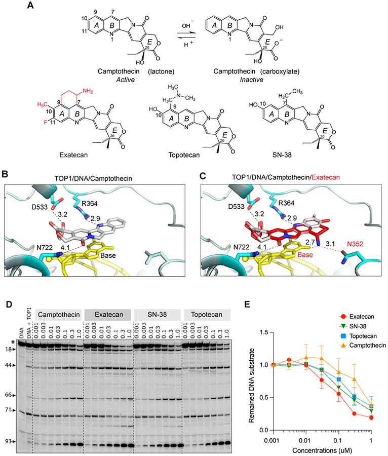
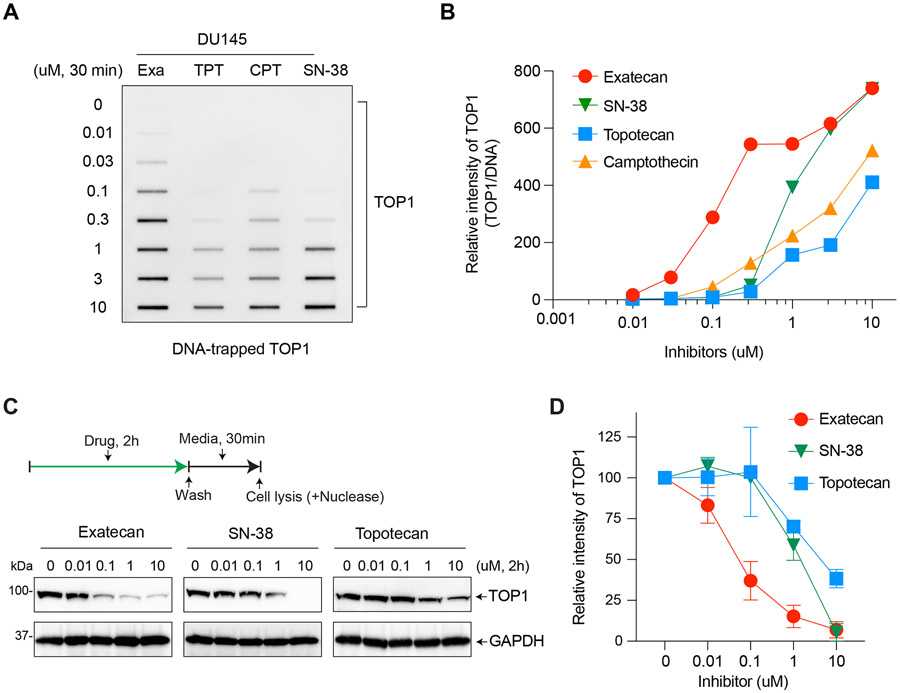
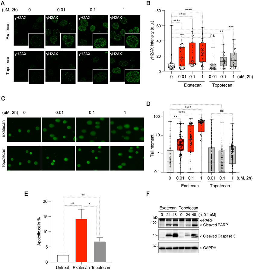
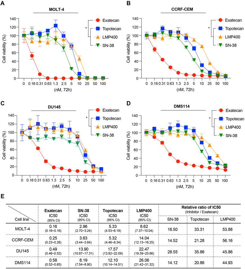
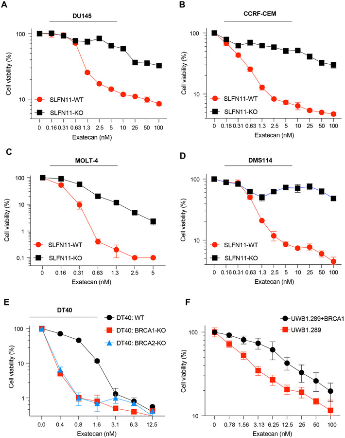
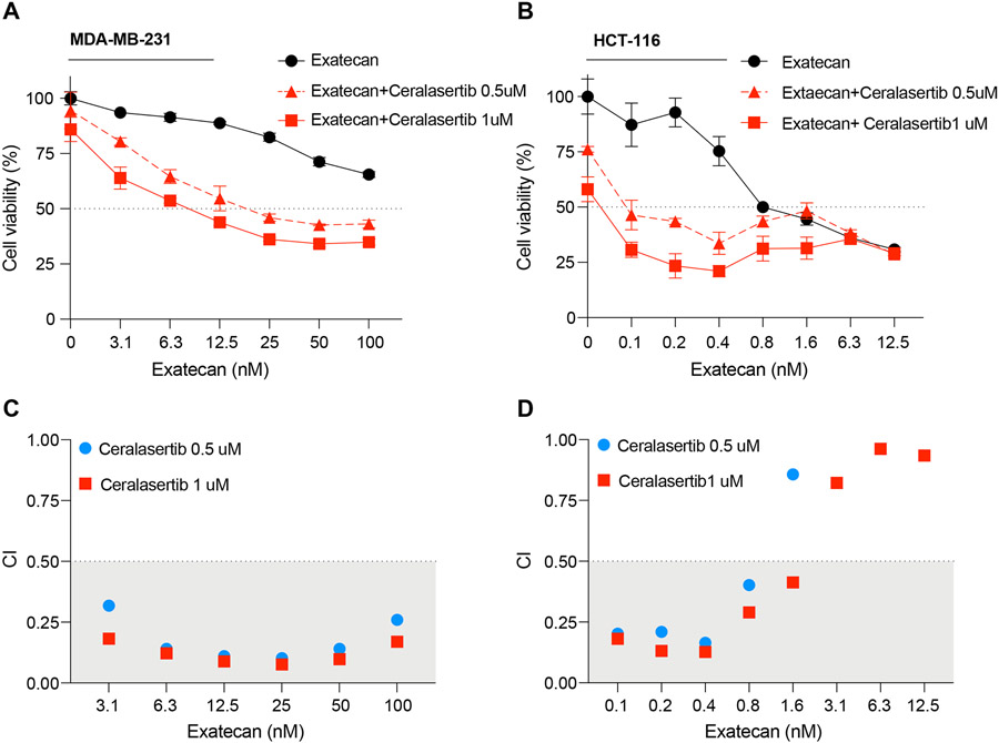
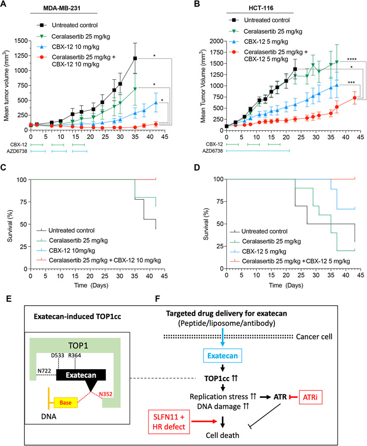
Exatecan and deruxtecan are antineoplastic camptothecin derivatives in development as tumor-targeted-delivery warheads in various formulations including peptides, liposomes, polyethylene glycol nanoparticles, and antibody-drug conjugates. Here, we report the molecular pharmacology of exatecan compared with the clinically approved topoisomerase I (TOP1) inhibitors and preclinical models for validating biomarkers and the combination of exatecan with ataxia telangiectasia and Rad3-related kinase (ATR) inhibitors. Modeling exatecan binding at the interface of a TOP1 cleavage complex suggests two novel molecular interactions with the flanking DNA base and the TOP1 residue N352, in addition to the three known interactions of camptothecins with the TOP1 residues R364, D533, and N722. Accordingly, exatecan showed much stronger TOP1 trapping, higher DNA damage, and apoptotic cell death than the classical TOP1 inhibitors used clinically. We demonstrate the value of SLFN11 expression and homologous recombination (HR) deficiency (HRD) as predictive biomarkers of response to exatecan. We also show that exatecan kills cancer cells synergistically with the clinical ATR inhibitor ceralasertib (AZD6738). To establish the translational potential of this combination, we tested CBX-12, a clinically developed pH-sensitive peptide-exatecan conjugate that selectively targets cancer cells and is currently in clinical trials. The combination of CBX-12 with ceralasertib significantly suppressed tumor growth in mouse xenografts. Collectively, our results demonstrate the potency of exatecan as a TOP1 inhibitor and its clinical potential in combination with ATR inhibitors, using SLFN11 and HRD as predictive biomarkers.


Conflict of interest statement

Conflict of Interest: R.S.B. is a co-founder and consultant for Cybrexa Therapeutics. UJ, YM, KA, YS, LKS, XY, YA, SG, KJ, VP, RKS, JVD, JCV, WSC, and YP declare no potential conflicts of interest.

Figures

Figure 1.
Figure 1.. Structural insights into the potent trapping of TOP1ccs by exatecan.
A. Chemical structures of CPT and its clinical derivatives (exatecan, topotecan, and SN-38). B. Representative view of CPT (light grey) bound to human TOP1 (cyan) and DNA (yellow) (PDB: 1T8I). In addition to base stacking, CPT makes 3 hydrogen bonds with TOP1 through D533, N722 and R364. The numbers indicate the distance of the bonds in Angstrom. C. Superposition of exatecan (red) and CPT (light grey) into the TOP1 (cyan)-DNA (yellow) structure. The 2 dotted lines (red) represent the potential additional hydrogen bonds of exatecan with the DNA base and N352 of TOP1. D. Comparative TOP1-mediated DNA cleavage (TOP1ccs) induced by exatecan and other TOP1 inhibitors. Recombinant TOP1 was incubated with 3'-end labeled 117 bp DNA oligo in the presence of the indicated drug concentrations. Fragmented DNA oligos were visualized on PAGE gel by using PhosphorImager (Molecular Dynamics). E. Quantitation of DNA substrates (*) as shown in panel D in duplicate experiments. The band intensity was determined by Image Quant software (Molecular Dynamics).
Figure 2.
Figure 2.. Exatecan leads to greater TOP1-DNA trapping than topotecan, SN-38 and CPT.
A. Detection of DNA-trapped TOP1 by exatecan and other TOP1 inhibitors. DU145 cells were treated with the indicated drug concentrations for 30 min. TOP1ccs were isolated by RADAR assay. B. Quantitation of TOP1ccs from panel A in a single experiment. The intensity of TOP1 was analyzed by ImageJ software and normalized to DNA loading. Data are plotted with GraphPad Prism 8. C. TOP1 degradation induced by exatecan. DU145 cells were incubated with the indicated TOP1 inhibitors for 2 h. Following TOP1 reversal for 30 min without inhibitors, TOP1 levels were determined by Western blotting. D. Quantification of total cellular TOP1 bands in duplicate experiments. Band intensity was analyzed using the ImageJ software and normalized to GAPDH used as a loading control.
Figure 3.
Figure 3.. DNA damage and cell death induced by exatecan.
A. Representative immunofluorescence images of γH2AX (green) in exatecan- or topotecan-treated DU145 cells. B. Intensity of γH2AX fluorescence (average per cell) for the experiment depicted in panels A (mean ± SEM, N = 50/each) ** p-value <0.002, ***p-value <0.0004, ****p-value <0.0001. a.u., arbitrary units. C. Representative images of comet analysis in DU145 cells treated with exatecan and topotecan. D. Quantitation of tail moments of experiments depicted in panel B (mean ± SEM, N = 100/each) are quantified with the Open Comet/ImageJ program. ** p-value <0.006, ****p-value <0.0001. E. Apoptotic cell death induced by exatecan and topotecan and measured by Annexin V/PI staining. * p-value <0.01, **p-value <0.005. F. Cleavage of PARP1 and caspase-3 in exatecan and topotecan treated cells measured by Western blotting.
Figure 4.
Figure 4.. Exatecan is the most potent TOP1 inhibitor
A~D. Cytotoxicity of clinical TOP1 inhibitors (Exatecan, SN-38, Topotecan, and LMP400) in MOLT-4, CCRF-CEM, DU145 and DMS114 cells. Cells were treated as indicated for 72 h and cell viability was measured by CellTiter-Glo assay. Error bars represent standard deviations in the triplicate. Statistical values were calculated using one-way ANOVA with Dunnett's multiple comparisons test. * p-value<0.03. E. IC50 values of the TOP1 inhibitors calculated by GraphPad Prism 8. The IC50 values represent the mean (nM) obtained from triplicate experiments in MOLT-4, CCRF-CEM, DMS114, and DU145 cells. CI, 95% confidence interval. Ratios indicate comparative IC50 values between exatecan and the other TOP1 inhibitors.
Figure 5.
Figure 5.. SLFN11-proficient and HR-deficient cells are preferentially vulnerable to exatecan.
A~D. Cytotoxicity of exatecan in the isogenic DU145, CCRF-CEM, MOLT-4, DMS114 and paired SLFN11 knock-out (KO) cells. Cells were treated as indicated for 72 h and cell viability was measured by CellTiter-Glo assay. E. Cytotoxicity of exatecan in the isogenic DT40 chicken B-cells and paired BRCA1/2 KO cells. Cells were treated as indicated for 72 h and cell viability was measured by CellTiter-Glo assay. F. Cytotoxicity of exatecan in UWB1.289 (carrying a BRCA1 mutation, BRCA1-null) and UWB1.289+BRCA1 cells. Cells were treated as indicated for 72 h, and cell viability was measured by e CellTiter-Glo assay. Error bars represent standard deviations in the triplicate.
Figure 6.
Figure 6.. Exatecan synergizes with the ATR inhibitor ceralasertib.
A-B. Cytotoxicity of combination treatments of exatecan with ATR inhibitor. Human breast cancer MDA-MB-231 and colon adenocarcinoma HCT116 cells were treated with the indicated concentrations of exatecan without or with ceralasertib (0.5 and 1 μM) for 72 h, and cell viability was measured by CellTiter-Glo assays. Error bars represent standard deviations in the triplicate. C-D. Combination index (CI) plots for the combinations exatecan and ceralasertib from data obtained from panels A and B. The CI values were calculated by using CompuSyn. Additive combination: 0.5<CI<1, and synergistic combinations: 0 <CI<0.5.
Figure 7.
Figure 7.. Antitumor activity of CBX-12 in human breast cancer and colon cancer xenografts and synergy with the ATR inhibitor ceralasertib.
A-B. Tumor suppression by CBX-12 without and with ceralasertib (AZD6738) in MDA-MB-231 and HCT-116 xenografts. MDA-MB-231 xenografts (A) were treated with CBX-12 intraperitoneally at 10 mg/kg once daily for 4 days, repeated weekly for 3 weeks. Ceralasertib was administered via oral gavage at 25 mg/kg once daily for 5 days, repeated weekly for 3 weeks. HCT-116 xenografts (B) were treated with CBX-12 intraperitoneally at 5 mg/kg once daily for 4 days, repeated weekly for 3 weeks. Ceralasertib doses were then administered via oral gavage at 25 mg/kg once daily for 21 days. Tumor volumes are shown as mean ± SEM (N = 10 mice for each group). Statistical values were calculated using one-way ANOVA with Dunnett's multiple comparisons test. * p-value<0.05, *** p-value<0.001, **** p-value<0.0001. C-D. Cell survival after drug treatments for the MDA-MB-231 (C) and HCT-116 (D) xenografts. E. Proposed model for potent TOP1cc trapping by exatecan. F. Therapeutic strategy and predictive biomarkers for targeted exatecan delivery. ATRi: ATR inhibitor, SLFN11: Schlafen 11 expression, HR: homologous recombination including BRCA1/2.

References

    1. Pommier Y, Marchand C. Interfacial inhibitors: targeting macromolecular complexes. Nat Rev Drug Discov 2011;11(1):25–36 doi 10.1038/nrd3404. - DOI - PMC - PubMed
    1. Thomas A, Pommier Y. Targeting Topoisomerase I in the Era of Precision Medicine. Clin Cancer Res 2019;25(22):6581–9 doi 10.1158/1078-0432.CCR-19-1089. - DOI - PMC - PubMed
    1. Kopetz S, Guthrie KA, Morris VK, Lenz HJ, Magliocco AM, Maru D, et al. Randomized Trial of Irinotecan and Cetuximab With or Without Vemurafenib in BRAF-Mutant Metastatic Colorectal Cancer (SWOG S1406). J Clin Oncol 2021;39(4):285–94 doi 10.1200/JCO.20.01994. - DOI - PMC - PubMed
    1. Mitsui I, Kumazawa E, Hirota Y, Aonuma M, Sugimori M, Ohsuki S, et al. A new water-soluble camptothecin derivative, DX-8951f, exhibits potent antitumor activity against human tumors in vitro and in vivo. Jpn J Cancer Res 1995;86(8):776–82 doi 10.1111/j.1349-7006.1995.tb02468.x. - DOI - PMC - PubMed
    1. Joto N, Ishii M, Minami M, Kuga H, Mitsui I, Tohgo A. DX-8951f, a water-soluble camptothecin analog, exhibits potent antitumor activity against a human lung cancer cell line and its SN-38-resistant variant. Int J Cancer 1997;72(4):680–6 doi 10.1002/(sici)1097-0215(19970807)72:4<680::aid-ijc21>3.0.co;2-e. - DOI - PubMed

Publication types

MeSH terms