. 2022 Apr 19;10(1):57.
doi: 10.1186/s40478-022-01359-y.

Impact of APOE genotype on prion-type propagation of tauopathy

Affiliations

Impact of APOE genotype on prion-type propagation of tauopathy

Tristan Williams et al. Acta Neuropathol Commun. .

Abstract

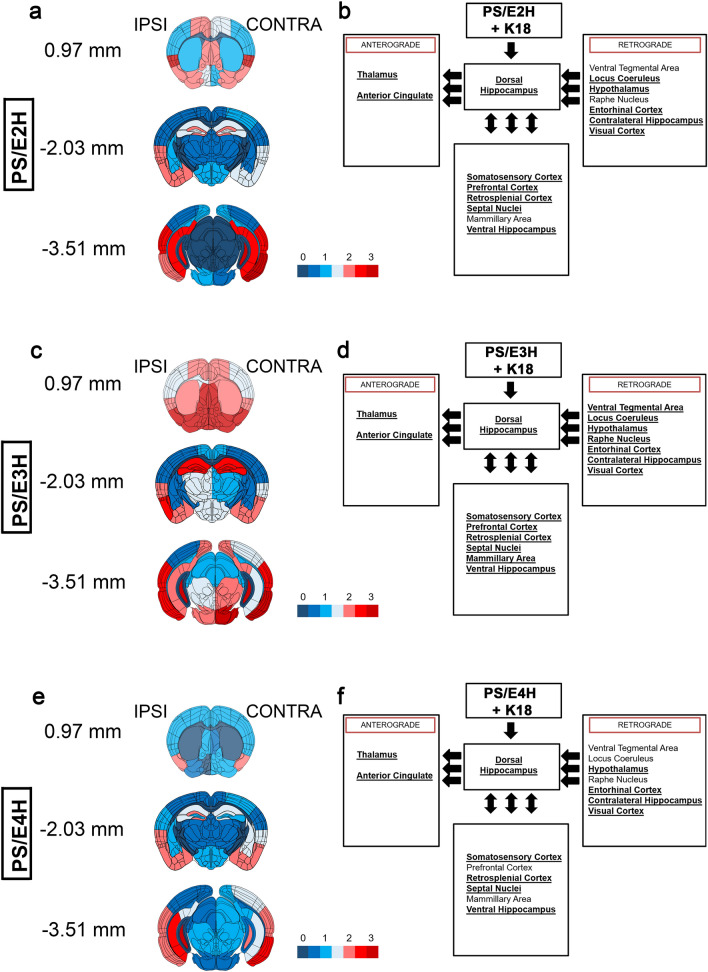
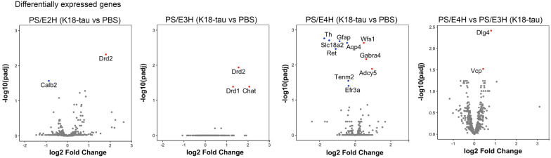
Apolipoprotein (APOE) is a major risk factor of Alzheimer's disease (AD), with the E2, E3 and E4 isoforms differentially regulating the burden of AD-associated neuropathologies, such as amyloid β and tau. In AD, pathological tau is thought to spread along neuroanatomic connections following a prion-like mechanism. To provide insights into whether APOE isoforms differentially regulate the prion properties of tau and determine trans-synaptic transmission of tauopathy, we have generated human P301S mutant tau transgenic mice (PS19) that carry human APOE (APOE2, APOE3 or APOE4) or mouse Apoe allele. Mice received intrahippocamal injections of preformed aggregates of K18-tau at young ages, which were analyzed 5 months post-inoculation. Compared to the parental PS19 mice with mouse Apoe alleles, PS19 mice expressing human APOE alleles generally responded to K18-tau seeding with more intense AT8 immunoreactive phosphorylated tau athology. APOE3 homozygous mice accumulated higher levels of AT8-reactive ptau and microgliosis relative to APOE2 or APOE4 homozygotes (E3 > E4~2). PS19 mice that were heterozygous for APOE3 showed similar results, albeit to a lesser degree. In the timeframe of our investigation, we did not observe significant induction of argentophilic or MC1-reactive neurofibrillary tau tangle in PS19 mice homozygous for human APOE. To our knowledge, this is the first comprehensive study in rodent models that provides neuropathological insights into the dose-dependent effect of APOE isoforms on phosphorylated tau pathology induced by recombinant tau prions.

Keywords: APOE; Phosphorylated tau; Prion; Tangle.


Conflict of interest statement

The authors declare no competing interests exist.

Figures

Fig. 1
Fig. 1
Accelerated induction of ptau pathology in PS/E3H mice seeded with K18-tau aggregates. K18-tau fibrils were injected into the left hippocampus of 2.5-month-old mice and aged for 5 months. 7.5-month-old mice were then analyzed using AT8 antibody. Representative images from the hippocampus and cortex of K18-tau aggregate injected hemisphere (ipsilateral, ‘IPSI’) and uninjected hemisphere (contralateral, ‘CONTRA’) showing ptau pathology in PS19 mice homozygous for APOE alleles (B6N2 generation) or PS19 mice carrying murine Apoe (a). Quantification of the antibody immunostaining is presented as % immunoreactivity in the cortex (Ctx) or hippocampus (Hpc) from ipsilateral and contralateral hemispheres around the injection site (b). Boxes in whole brain panel indicate selected areas used for high power zoomed panels. n = 9–12 mice/group. 1-way ANOVA *p < 0.05, **p < 0.01, ***p < 0.001, ****p < 0.0001. Scale bar: 3 mm (whole brain); 100 µm (hippocampus and cortex)
Fig. 2
Fig. 2
Relative abundance of ptau in hippocampus and neuroanatomically connected brain regions in K18-tau aggregate seeded PS19 mice carrying human APOE alleles. Semi-quantitative analyses of ptau transmission patterns along neuroanatomic pathways represented by AT8 immunostaining is shown from K18-tau aggregate seeded PS19 mice homozygous for human APOE alleles (B6N2 generation). Pathology severity was assigned scores on a scale of 0 (no pathology) to 3 (high pathology) and color-coded onto heat maps (a, c, e). The injected hemisphere (ipsilateral, ‘IPSI’) is shown on the left and non-injected (contralateral, ‘CONTRA’) hemisphere is depicted on the right for each heat map. Three coronal planes were examined at bregma locations of 0.97, − 2.03 (site of injection), and − 3.51 mm. Boxed diagrams on the right show different brain regions neuroanatomically connected to the dorsal hippocampus via either anterograde or retrograde pathways (left pointing arrows) or both pathways (double-headed arrows) (b, d, f). Brain regions showing AT8 immunoreactivity are indicated by bold and underlined text in the boxes (b, d, f). See also Additional file 1: Fig. S1. n = 3–4 mice/group
Fig. 3
Fig. 3
Differential gene expression patterns in K18-tau aggregate seeded PS19 mice homozygous for APOE. nCounter Neuropathology panel was used to assess differential gene expression patterns in K18-tau aggregate seeded PS/E2H, PS/E3H and PS/E4H mice relative to age- and genotype-matched PBS injected mice (left three panels). Genes differentially regulated in K18-tau aggregate seeded PS/E4H mice relative to PS/E3H mice is shown on the right panel. p values adjusted for multiple testing; FDR = 0.05. n = 3–4 mice/group
Fig. 4
Fig. 4
Induction of gliosis in K18-tau seeded PS19 mice homozygous for APOE. K18-tau fibrils were injected into the left hippocampus of 2.5-month-old mice and aged for 5 months. 7.5-month-old mice were analyzed for Iba-1 immunoreactive microgliosis (a, b) and GFAP-immunoreactive astrogliosis (c, d). Representative images from the hippocampus and cortex of K18-tau aggregate injected hemisphere (ipsilateral, ‘IPSI’) and uninjected hemisphere (contralateral, ‘CONTRA’) are shown (a, c). Quantification of the Iba-1 and GFAP immunostaining in 7.5-month-old mice is presented as % immunoreactivity in the cortex (Ctx) or hippocampus (Hpc) (b, d). Boxes in whole brain panel indicate selected areas used for high power zoomed panels. n = 9–12 mice/group. 1-way ANOVA *p < 0.05, **p < 0.01, ***p < 0.001. Scale bar: 3 mm (whole brain); 100 µm (hpc and ctx)
Fig. 5
Fig. 5
Comparative analysis of intrahippocampal pathology in PS19 mice carrying human APOE alleles or murine Apoe. Graphical representation of AT8 immunoreactivity levels in bigenic PS19 mice carrying human APOE alleles from the B6N2 and B6N1 generations and parental PS19 mice carrying mouse Apoe allele. N1 (B6N1) and N2 (B6N2) refer to two subsequent generations with slightly different mouse genetic backgrounds. AT8 burden from PBS injected mice (a, c, e) and K18-tau aggregate seeded mice (b, d, f) shown from three different PS19xAPOE cohorts. Data originally shown in Fig. 2, S8 and S9

References

    1. Goedert M, Eisenberg DS, Crowther RA. Propagation of Tau aggregates and neurodegeneration. Annu Rev Neurosci. 2017;40:189–210. doi: 10.1146/annurev-neuro-072116-031153. - DOI - PubMed
    1. Brettschneider J, et al. Spreading of pathology in neurodegenerative diseases: a focus on human studies. Nat Rev Neurosci. 2015;16(2):109–120. doi: 10.1038/nrn3887. - DOI - PMC - PubMed
    1. Yamazaki Y, et al. Apolipoprotein E and Alzheimer disease: pathobiology and targeting strategies. Nat Rev Neurol. 2019;15(9):501–518. doi: 10.1038/s41582-019-0228-7. - DOI - PMC - PubMed
    1. Long JM, Holtzman DM. Alzheimer disease: an update on pathobiology and treatment strategies. Cell. 2019;179(2):312–339. doi: 10.1016/j.cell.2019.09.001. - DOI - PMC - PubMed
    1. Lewandowski CT, Maldonado Weng J, LaDu MJ. Alzheimer's disease pathology in APOE transgenic mouse models: the who, what, when, where, why, and how. Neurobiol Dis. 2020;139:104811. doi: 10.1016/j.nbd.2020.104811. - DOI - PMC - PubMed