Review
. 2022 Jul 8:45:515-531.
doi: 10.1146/annurev-neuro-111020-091953. Epub 2022 Apr 19.

Human Cerebellar Development and Transcriptomics: Implications for Neurodevelopmental Disorders

Affiliations
Review

Human Cerebellar Development and Transcriptomics: Implications for Neurodevelopmental Disorders

Parthiv Haldipur et al. Annu Rev Neurosci. .

Abstract

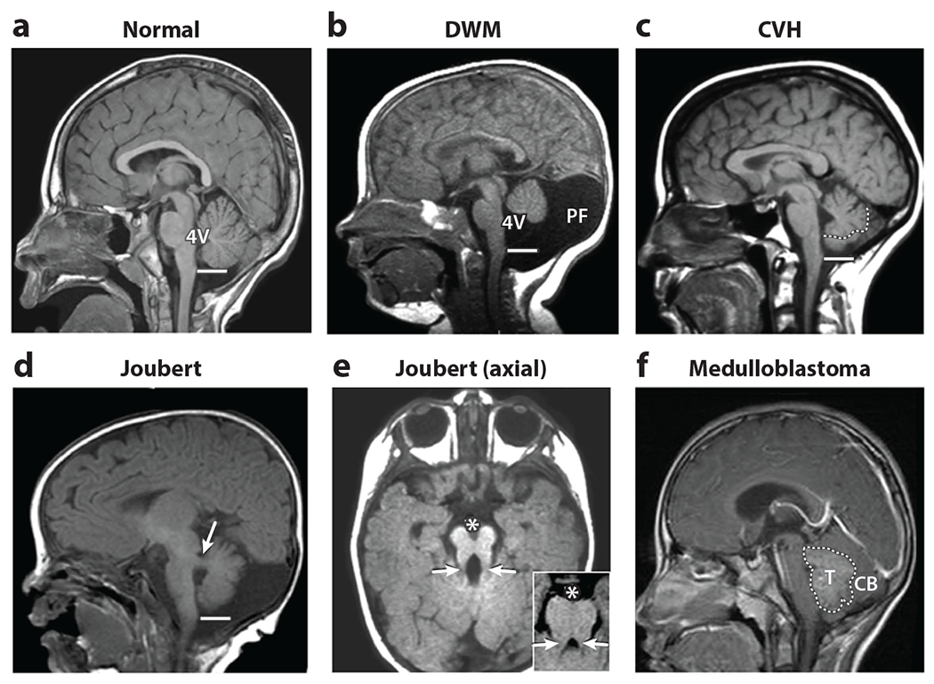
Developmental abnormalities of the cerebellum are among the most recognized structural brain malformations in human prenatal imaging. Yet reliable information regarding their cause in humans is sparse, and few outcome studies are available to inform prognosis. We know very little about human cerebellar development, in stark contrast to the wealth of knowledge from decades of research on cerebellar developmental biology of model organisms, especially mice. Recent studies show that multiple aspects of human cerebellar development significantly differ from mice and even rhesus macaques, a nonhuman primate. These discoveries challenge many current mouse-centric models of normal human cerebellar development and models regarding the pathogenesis of several neurodevelopmental phenotypes affecting the cerebellum, including Dandy-Walker malformation and medulloblastoma. Since we cannot model what we do not know, additional normative and pathological human developmental data are essential, and new models are needed.

Keywords: genetics; histology; malformation; neurogenesis; rhombic lip; ventricular zone.


Figures

Figure 1
Figure 1
Neuroimaging findings define human cerebellar malformations and anomalies. (a) A normal midline sagittal T1-weighted image demonstrates the exquisite structure of the cerebellar vermis extending to the obex of the brainstem (line) adjacent to the fourth ventricle (4V). (b) Dandy-Walker malformation (DWM) is characterized by a small cerebellar vermis, which is rotated away from the brainstem, resulting in an enlarged 4V and space at the back of the skull [posterior fossa (PF)]. (c) In cerebellar vermis hypoplasia (CVH), the cerebellar vermis is hypoplastic without an enlarged 4V or PF (the vermis is outlined by a dotted line to distinguish it from the lateral cerebellar hemispheres, which collapsed to occupy medial space). (d) Joubert syndrome is characterized by a small cerebellar vermis, which also fails to extend to the obex, together with thickened and abnormally positioned cerebellar peduncles (white arrow) in this paramedial sagittal image. (e) In axial images, these abnormal peduncles (white arrows), together with a large interpeduncular fissure (denoted by *), result in a molar tooth sign (inset), never seen in normal individuals. (f) In this example of medulloblastoma, a tumor mass (T) obscures the normal cerebellar (CB) structure. Figure adapted from Chizhikov & Millen (2013).
Figure 2
Figure 2
Timeline of mouse and human cerebellar development. Cerebellar neurogenesis in the mouse and human is driven by three progenitor zones: two primary zones, namely the VZ and RL, and one secondary zone, the EGL. Development of each cell type is indicated by a unique colored line. In mice, cerebellar development takes place over a period of approximately 30–35 days, with a significant portion occurring in the postnatal period. In humans, cerebellar development is protracted, taking place over 2–3 years, beginning approximately 30 days post-conception, with all major developmental events taking place in utero. Peak progenitor proliferation is indicated by thickened dashed lines. In both species, VZ- and RL-driven growth precedes an EGL-driven increase in cerebellar volume and foliation. However, unlike in the mouse, where RL presence is transient, in humans, the RL is spatiotemporally expanded and promotes growth and maintenance of the posterior lobe throughout gestation. Abbreviations: e, embryonic day; EGL, external granule layer; P, postnatal day; PC, Purkinje cell; pcw, postconceptional week; PN, postnatal; RL, rhombic lip; SVZ, subventricular zone; VZ, ventricular zone.
Figure 3
Figure 3
Genetic and prenatal factors cause cerebellar malformations. The cumulative genetic and prenatal injury rates associated with cerebellar malformations are depicted as stacked bar graphs. The prenatal injury rate is constant among cerebellar malformation types, but genetic rates vary among cerebellar malformation diagnoses. Exome sequencing is nondiagnostic for Dandy-Walker malformation among the patients tested. Here, cerebellar hypoplasia is used as a catchall term for cerebellar malformation types that are not Dandy-Walker malformation. Data are based on published exome sequencing, review of prenatal history, and review of neuroimaging (Aldinger et al. 2019).

References

    1. Abraham H, Tornoczky T, Kosztolanyi G, Seress L. 2001. Cell formation in the cortical layers of the developing human cerebellum. Int. J. Dev. Neurosci 19:53–62 - PubMed
    1. Aguilar A, Meunier A, Strehl L, Martinovic J, Bonniere M, et al. 2012. Analysis of human samples reveals impaired SHH-dependent cerebellar development in Joubert syndrome/Meckel syndrome. PNAS 109:16951–56 - PMC - PubMed
    1. Aldinger KA, Dempsey JC,Tully HM, Grout ME, Mehaffey MG, et al. 2018. Rhombencephalosynapsis: fused cerebellum, confused geneticists. Am. J. Med. Genet. C Semin. Med. Genet 178:432–39 - PMC - PubMed
    1. Aldinger KA, Doherty D. 2016. The genetics of cerebellar malformations. Semin. Fetal Neonatal. Med 21:321–32 - PMC - PubMed
    1. Aldinger KA, Thomson Z, Phelps IG, Haldipur P, Deng M, et al. 2021. Spatial and cell type transcriptional landscape of human cerebellar development. Nat. Neurosci 24:1163–75 - PMC - PubMed

Publication types

LinkOut - more resources