Review
. 2022 Apr;12(4):210318.
doi: 10.1098/rsob.210318. Epub 2022 Apr 20.

The role of procoagulant phospholipids on the surface of circulating blood cells in thrombosis and haemostasis

Affiliations
Review

The role of procoagulant phospholipids on the surface of circulating blood cells in thrombosis and haemostasis

Majd B Protty et al. Open Biol. 2022 Apr.

Abstract

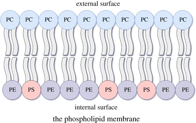
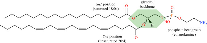
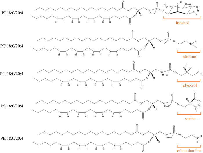
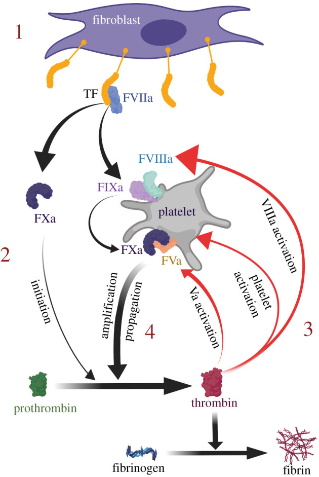
Phospholipids (PLs) are found in all cell types and are required for structural support and cell activation signalling pathways. In resting cells, PLs are asymmetrically distributed throughout the plasma membrane with native procoagulant aminophospholipids (aPLs) being actively maintained in the inner leaflet of the membrane. Upon platelet activation, aPLs rapidly externalize to the outer leaflet and are essential for supporting the coagulation cascade by providing binding sites for factors in the cell-based model. More recent work has uncovered a role for enzymatically oxidized PLs (eoxPLs) in facilitating coagulation, working in concert with native aPLs. Despite this, the role of aPLs and eoxPLs in thrombo-inflammatory conditions, such as arterial and venous thrombosis, has not been fully elucidated. In this review, we describe the biochemical structures, distribution and regulation of aPL externalization and summarize the literature on eoxPL generation in circulating blood cells. We focus on the currently understood role of these lipids in mediating coagulation reactions in vitro, in vivo and in human thrombotic disease. Finally, we highlight gaps in our understanding in how these lipids vary in health and disease, which may place them as future therapeutic targets for the management of thrombo-inflammatory conditions.

Keywords: coagulation; lipidomics; phospholipids; thrombosis.


Conflict of interest statement

All authors have no relevant affiliations or financial involvement with any organization or entity with a financial interest in or financial conflict with the subject matter or materials discussed in the manuscript.

Figures

Figure 1.
Figure 1.
A simplified illustration of native phospholipid (PL) plasma membrane relevant to coagulation. Polarized PLs make up the membrane bilayer of all mammalian cells with phosphate head groups facing the aqueous phase and hydrophobic fatty acids facing the core. Other lipids and proteins line the membrane, which may also influence procoagulant membrane activity (e.g. sphingomyelin), but are outside the scope of this review and therefore not shown in this figure. PS, phosphatidylserine; PE, phosphatidylethanolamine; PC: phosphatidylcholine.
Figure 2.
Figure 2.
Example of a phospholipid molecule demonstrating the sn1/sn2/headgroup positions on the glycerol backbone. In this example, 1-stearoyl-2-arachidonyl-phosphatidylethanolamine, or PE 18:0a/20:4, is demonstrated with the glycerol backbone highlighted in a green polygon. Structures drawn with the aid of tools on LIPID MAPS (www.lipidmaps.org).
Figure 3.
Figure 3.
Phospholipid classes and chemical structures highlighting the phosphate head groups. In these images, the sn1 fatty acid is stearic acid (FA 18:0) and the sn2 fatty acid is arachidonic acid (FA 20:4). The structures of the five head groups can also be seen (PI, phosphatidylinositol; PC, phosphatidylcholine; PG, phosphatidylglycerol; PS, phosphatidylserine; PE, phosphatidylethanolamine). Structures drawn with the aid of tools on the LIPID MAPS resource (www.lipidmaps.org).
Figure 4.
Figure 4.
The coagulation system (cell-based model). Activation of coagulation is driven by tissue factor (TF) expressing cells in the subendothelial space (1). The TF:FVIIa complex activation of FX to FXa and FIX to FIXa is termed the ‘initiation phase’, which generates small amounts of thrombin (2). This is sufficient to activate FV to FVa and FVIII to FVIIIa, leading to the formation of FIXa:FVIIIa and FXa:FVa complexes on the platelet PL surface (3). These complexes lead to the formation of more FXa and more thrombin, respectively, as part of the ‘amplification phase’. More thrombin leads to more activated platelets and coagulation factors locally, creating a thrombin-forming ‘propagation phase’ loop (4) which leads to the formation of fibrin.
Figure 5.
Figure 5.
The typical oxylipin and enzymatically oxidized phospholipid pathway in circulating blood cells. Membrane phospholipids can be cleaved by phospholipase A2 (PLA2) into polyunsaturated fatty acids (PUFAs), such as arachidonic acid (FA 20:4). These PUFAs are oxygenated via the action of cyclooxygenase (COX) or lipoxygenase (LOX) enzymes to generate oxylipins, which are referred to as ‘eicosanoids’ if generated from 20-carbon PUFAs such as arachidonic acid (AA). Some oxylipins may be re-esterified back to the membrane to form enzymatically oxidized phospholipids. The ‘n-’ prefix denotes the enzyme isoforms which are responsible for generating oxylipin positional isomers at the corresponding ‘n-’ carbon on AA (e.g. 12-LOX in platelets generating 12-hydroxyeicosatetraenoic acids, or 12-HETE). HPETE, hydroperoxyeicosatetraenoic acid; LT, leukotriene.
Figure 6.
Figure 6.
The mechanism of action of LOXs. Hydrogen atom abstraction from the PUFA substrate proceeds through a proton-coupled electron transfer (PCET) mechanism, with simultaneous transfer of electrons and protons via a concerted mechanism. Subsequently, radical rearrangement takes place with stereospecific dioxygen insertion. This is followed by a further PCET step to generate a PUFA hydroperoxide, which can then be reduced by glutathione peroxidase (GPX) enzymes to the hydroxide forms. In the case of AA, the hydroperoxides are known as HPETEs, hydroxides are known as HETEs and the position of the oxygen insertion (forming (S) stereoisomers) is dictated by the cell-specific LOX isoform. LOX, lipoxygenase, PUFA, polyunsaturated fatty acid; AA, arachidonic acid; HPETE, hydroperoxyeicosatetraenoic acid; HETE, hydroxyeicosatetraenoic acid. Adapted from Hajeyah et al. [58].
Figure 7.
Figure 7.
Reaction mechanism of cyclooxygenase (COX) and the generation of 11-HETE and 15-HETE as by-products of the dioxygenase reactions. Arachidonic acid (AA) undergoes hydrogen atom abstraction followed by radical rearrangement to allow for the insertion of a dioxygen molecule. The resultant product undergoes endoperoxide formation and cyclization to ultimately generate prostaglandin H2 (PGH2)—the intermediary product of COX-derived prostaglandins and thromboxanes. The dioxygenated AA may also undergo reduction instead of cyclization, leading to the generation of 11(R)-HPETE and subsequent reduction by glutathione peroxidase (GPX) enzymes to 11(R)-HETE. Alternative radical rearrangement to carbon 15 (instead of 11) can lead to the generation of 15(R)-HETE downstream of COX metabolism of AA. HPETE, hydroperoxyeicosatetraenoic acid; HETE, hydroxyeicosatetraenoic acid. Adapted from Hajeyah et al. [58].
Figure 8.
Figure 8.
The enzymatic pathways of eoxPL biosynthesis. This figure depicts the three different pathways known to date for eoxPL generation: (1) PLA2 hydrolyses membrane PLs, releasing the sn2 PUFA, which is then oxygenated by COX-11/2, LOXs or cytochrome p450 enzymes to generate oxylipins (oxPUFA). These undergo the addition of coenzyme A (CoA) via acyl-CoA synthase (ACSL), prior to being re-esterified into a lysophospholipid (lysoPL) by any one of a series of sn2 acyltransferases (also known as LPLATs). (2) The phospholipase PLA1 hydrolyses membrane PLs to release 2-PUFA-lysophospholipids, which may be oxygenated by COX-2, 12S-LOX and 15-LOX prior to being re-esterified with a fatty acid CoA (FA-CoA). (3) Unique to 15-LOX is the ability to directly oxygenate membrane PL to form eoxPL.

References

    1. O'Donnell VB, Murphy RC, Watson SP. 2014. Platelet lipidomics: modern day perspective on lipid discovery and characterization in platelets. Circ. Res. 114, 1185-1203. ( 10.1161/CIRCRESAHA.114.301597) - DOI - PMC - PubMed
    1. Burdge GC, Calder PC. 2015. Introduction to fatty acids and lipids. World Rev. Nutr. Diet 112, 1-16. ( 10.1159/000365423) - DOI - PubMed
    1. Vance DE, Vance JE. 2008. Biochemistry of lipids, lipoproteins, and membranes, 5th edn. Amsterdam, The Netherlands: Elsevier.
    1. Liebisch G, Vizcaíno JA, Köfeler H, Trötzmüller M, Griffiths WJ, Schmitz G, Spener F, Wakelam MJ. 2013. Shorthand notation for lipid structures derived from mass spectrometry. J. Lipid Res. 54, 1523-1530. ( 10.1194/jlr.M033506) - DOI - PMC - PubMed
    1. O'Donnell VB, Rossjohn J, Wakelam MJ. 2018. Phospholipid signaling in innate immune cells. J. Clin. Invest 128, 2670-2679. ( 10.1172/JCI97944) - DOI - PMC - PubMed

Publication types