. 2022 May 18;42(20):4215-4228.
doi: 10.1523/JNEUROSCI.1910-21.2022. Epub 2022 Apr 19.

Chronic Cortical Inflammation, Cognitive Impairment, and Immune Reactivity Associated with Diffuse Brain Injury Are Ameliorated by Forced Turnover of Microglia

Affiliations

Chronic Cortical Inflammation, Cognitive Impairment, and Immune Reactivity Associated with Diffuse Brain Injury Are Ameliorated by Forced Turnover of Microglia

Chelsea E Bray et al. J Neurosci. .

Abstract

Traumatic brain injury (TBI) is associated with an increased risk of cognitive, psychiatric, and neurodegenerative complications that may develop after injury. Increased microglial reactivity following TBI may underlie chronic neuroinflammation, neuropathology, and exaggerated responses to immune challenges. Therefore, the goal of this study was to force turnover of trauma-associated microglia that develop after diffuse TBI and determine whether this alleviated chronic inflammation, improved functional recovery and attenuated reduced immune reactivity to lipopolysaccharide (LPS) challenge. Male mice received a midline fluid percussion injury (mFPI) and 7 d later were subjected to a forced microglia turnover paradigm using CSF1R antagonism (PLX5622). At 30 d postinjury (dpi), cortical gene expression, dendritic complexity, myelin content, neuronal connectivity, cognition, and immune reactivity were assessed. Myriad neuropathology-related genes were increased 30 dpi in the cortex, and 90% of these gene changes were reversed by microglial turnover. Reduced neuronal connectivity was evident 30 dpi and these deficits were attenuated by microglial turnover. TBI-associated dendritic remodeling and myelin alterations, however, remained 30 dpi independent of microglial turnover. In assessments of functional recovery, increased depressive-like behavior, and cognitive impairment 30 dpi were ameliorated by microglia turnover. To investigate microglial priming and reactivity 30 dpi, mice were injected intraperitoneally with LPS. This immune challenge caused prolonged lethargy, sickness behavior, and microglial reactivity in the TBI mice. These extended complications with LPS in TBI mice were prevented by microglia turnover. Collectively, microglial turnover 7 dpi alleviated behavioral and cognitive impairments associated with microglial priming and immune reactivity 30 dpi.SIGNIFICANCE STATEMENT A striking feature of traumatic brain injury (TBI), even mild injuries, is that over 70% of individuals have long-term neuropsychiatric complications. Chronic inflammatory processes are implicated in the pathology of these complications and these issues can be exaggerated by immune challenge. Therefore, our goal was to force the turnover of microglia 7 d after TBI. This subacute 7 d postinjury (dpi) time point is a critical transitional period in the shift toward chronic inflammatory processes and microglia priming. This forced microglia turnover intervention in mice attenuated the deficits in behavior and cognition 30 dpi. Moreover, microglia priming and immune reactivity after TBI were also reduced with microglia turnover. Therefore, microglia represent therapeutic targets after TBI to reduce persistent neuroinflammation and improve recovery.

Keywords: brain injury; cognition; forced turnover; inflammation; lipopolysaccharide; microglia.


Figures

Figure 1.
Figure 1.
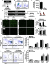
PLX5622-mediated elimination and repopulation of brain myeloid cells. A, Adult male C57BL/6 mice were provided diets formulated with either vehicle or a CSF1R antagonist (PLX5622; PLX) at for 7d. Brain myeloid cells were enriched by Percoll isolation, and the percentage of microglia were determined in the brain (n = 3). B, Representative bivariate dot plots of CD11b/CD45 labeling of Percoll-enriched cells. C, Number of microglia (CD11b+/CD45low) in the brain, normalized to control counting beads. D, Adult male C57BL/6 mice were subjected to mFPI (TBI) or left as uninjured controls (CON). At 7 dpi, mice were provided diets formulated with either vehicle or a CSF1R antagonist (PLX5622; PLX) for 7 d and then returned to standard chow 14 dpi. At 30 dpi (16 d of microglia repopulation), microglial histology (Iba-1+ labeling) was determined. E, Representative images of Iba-1+ labeling in the cortex 14 dpi (PLX-Depletion) and 30 dpi (PLX-Repop). Percent area of Iba-1+ microglia (F) 14 dpi and (G) 30 dpi (n = 6). In a parallel experiment using this design, the percentage of cells in the bone marrow and spleen were determined at the 30-d endpoint (16 d of repopulation). H, Representative bivariate dot plots of Ly6C/Ly6G labeling of Terr119/Cd11b+ cells isolated from the bone marrow after 16 d of repopulation. I, Percentage of monocytes, granulocytes, B-cells, and other lymphocytes in the bone marrow after 14 d of repopulation (n = 6). J, Representative bivariate dot plots of Ly6C/Ly6G labeling of Ter119/Cd11b+ cells isolated from the spleen after 16 d of repopulation. K, Percentage of monocytes, granulocytes, B-cells, and T-cells in the spleen after 16 d of repopulation (n = 6). Bars represent the mean ± SEM. Means with * are significantly different from control (p < 0.05).
Figure 2.
Figure 2.
Forced microglial turnover attenuated persistent neuropathology-related mRNA expression in the cortex 30 dpi. A, Adult male C57BL/6 mice were subjected to mFPI (TBI) or left as uninjured controls (CON). At 7 dpi, mice were provided diets formulated with either vehicle or a CSF1R antagonist (PLX5622; PLX) for 7d and then returned to standard chow 14 dpi. At 30 dpi (16 d of microglia repopulation), cortical mRNA expression (NanoString neuropathology panel) in the cortex was determined (n = 6). B, Heatmap shows the relative z score of mRNA that was differentially expressed by TB1 and influenced by PLX-Repop. Means with * are significantly different from CON-Veh (p < 0.05) and means with ^ are significantly different from both CON-Veh and TB1-Veh (p < 0.05). C, Pie chart represents genes unaffected (5 genes) or restored (48 genes) following microglial turnover after TBI. Select genes are shown and annotated if the gene is predominately expressed by a primary cell-type (Brain RNAseq, www.brainrnaseq.org). IPA of (D) significant upstream regulators (composite z score, p < 0.05) and (E) significant canonical pathways (activation z score, p < 0.05) of the differentially expressed genes based on their respective z score are shown.
Figure 3.
Figure 3.
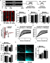
Forced turnover of microglia attenuated deficits in neuronal connectivity 30 dpi. A, Adult male C57BL/6 mice were subjected to mFPI (TBI) or left as uninjured controls (CON). At 7 dpi, mice were provided diets formulated with either vehicle or a CSF1R antagonist (PLX5622) for 7 d and then returned to their standard rodent chow at 14 dpi. At 30 dpi (16 d of microglia repopulation), mice were killed, and tissues were collected for analysis of dendritic spines, MBP, or CAPs. B, Representative cortical images of DiI+ dendrites 30 dpi from each treatment group are shown (n = 6). Bar graphs represent (C) dendritic spine volume, (D) dendritic spine area, and (E) dendritic spine density. F, CAPs were stimulated and measured from ex vivo preparation of the corpus callosum (n = 4) and representative N1 and N2 tracings of CAP from control and TB1 mice 30 dpi are shown. Graphs represent average recording amplitude across a range of stimulus intensities for (G) N1 and (H) N2. AUC determined for the (I) N1 and J) N2 amplitudes. K, Representative cortical images of MBP+ labeling 30 dpi from each treatment group (n = 6). L, Percent MBP+ labeling in cortical sections. Graphs represent mean ± SEM. Means with * are significantly different from CON-Veh (p < 0.05) and means with are significantly different from both CON-Veh and PLX-Repop (p < 0.05).
Figure 4.
Figure 4.
Cognitive deficits and depressive-like behavior 30 dpi were ameliorated by microglial turnover. A, Adult male C57BL/6 mice were subjected to mFPI (TBI) or left as uninjured controls. At 7 dpi, mice were provided diets formulated with either vehicle or a CSF1R antagonist (PLX5622) for 7 d, and then returned to standard rodent chow at 14 dpi. At 30–31 dpi (16 d of microglia repopulation), depressive-like behavior (n = 9) and cognition (n = 12) were assessed. B, Percentage of time immobile in the TST at 30 dpi. In a separate cohort, mice were used in the NOL and NOR tests. For NOL at 30 dpi, (C) time spent exploring the arena, (D) percentage of time spent investigating the object location, and (E) discrimination index for the object location were determined. For NOR at 31 dpi, (F) time spent exploring the arena, (G) percent of time spent investigating the novel object, and (H) discrimination index for the novel object were determined. Graphs represent mean ± SEM. Means with * are significantly different from CON-Veh (p < 0.05).
Figure 5.
Figure 5.
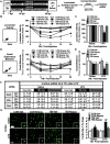
Immune reactivity 30 dpi was reduced by forced turnover of microglia. A, Adult male C57BL/6 mice were subjected to mFPI (TBI) or left as uninjured controls. At 7 dpi, mice were provided diets formulated with either vehicle or a CSF1R antagonist (PLX5622) for 7d, and then returned to their standard rodent chow at 14 dpi. At 30 dpi (16 d of microglia repopulation), mice received an intraperitoneal injection of either saline or LPS (0.5 mg/kg) and locomotor activity and social exploratory behavior were assessed 0, 4, 8, 12, and 24 h later (n = 9). At 24 (n = 6) and 72 h (n = 8) after LPS, a coronal section was collected for mRNA analyses. B, Time spent mobile in the open field over the 24-h time course. C, Time spent mobile in the open field at 24 h postinjection. D, Social exploratory behavior with a novel juvenile over the 24-h time course postinjection. E, Social behavior with a novel juvenile at 24 h postinjection. F, mRNA levels of Ccl2, Il1b, and Tlr4 were determined in a coronal brain section 24 and 72 h postinjection. G, Representative images of Iba-1+ labeling in the cortex 72 h postinjection. H, Percent area of Iba-1+ microglia labeling in the cortex (n = 5). Bars represent mean ± SEM. Means with * are significantly different from CON-Veh (p < 0.05) and means with are significantly different from both CON-Veh and PLX-Repop (p < 0.05).

References

    1. Abdullah A, Zhang M, Frugier T, Bedoui S, Taylor JM, Crack PJ (2018) STING-mediated type-I interferons contribute to the neuroinflammatory process and detrimental effects following traumatic brain injury. J Neuroinflammation 15:323. 10.1186/s12974-018-1354-7 - DOI - PMC - PubMed
    1. Antunes M, Biala G (2012) The novel object recognition memory: neurobiology, test procedure, and its modifications. Cogn Process 13:93–110. 10.1007/s10339-011-0430-z - DOI - PMC - PubMed
    1. Bachstetter AD, Rowe RK, Kaneko M, Goulding D, Lifshitz J, Van Eldik LJ (2013) The p38alpha MAPK regulates microglial responsiveness to diffuse traumatic brain injury. J Neurosci 33:6143–6153. 10.1523/JNEUROSCI.5399-12.2013 - DOI - PMC - PubMed
    1. Bachstetter AD, Van Eldik LJ, Schmitt FA, Neltner JH, Ighodaro ET, Webster SJ, Patel E, Abner EL, Kryscio RJ, Nelson PT (2015) Disease-related microglia heterogeneity in the hippocampus of Alzheimer's disease, dementia with Lewy bodies, and hippocampal sclerosis of aging. Acta Neuropathol Commun 3:32. 10.1186/s40478-015-0209-z - DOI - PMC - PubMed
    1. Barrett JP, Henry RJ, Shirey KA, Doran SJ, Makarevich OD, Ritzel RM, Meadows VA, Vogel SN, Faden AI, Stoica BA, Loane DJ (2020) Interferon-beta plays a detrimental role in experimental traumatic brain injury by enhancing neuroinflammation that drives chronic neurodegeneration. J Neurosci 40:2357–2370. 10.1523/JNEUROSCI.2516-19.2020 - DOI - PMC - PubMed

Publication types

Substances