. 2022 Apr 19;5(8):e202201421.
doi: 10.26508/lsa.202201421. Print 2022 Aug.

LuminoCell: a versatile and affordable platform for real-time monitoring of luciferase-based reporters

Affiliations

LuminoCell: a versatile and affordable platform for real-time monitoring of luciferase-based reporters

Kamila Weissová et al. Life Sci Alliance. .

Abstract

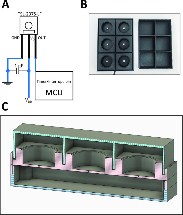
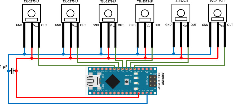
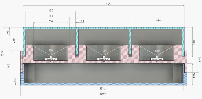
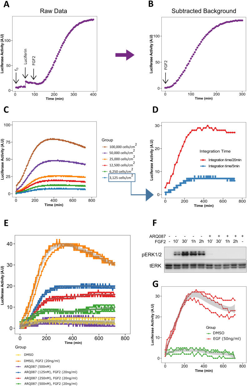
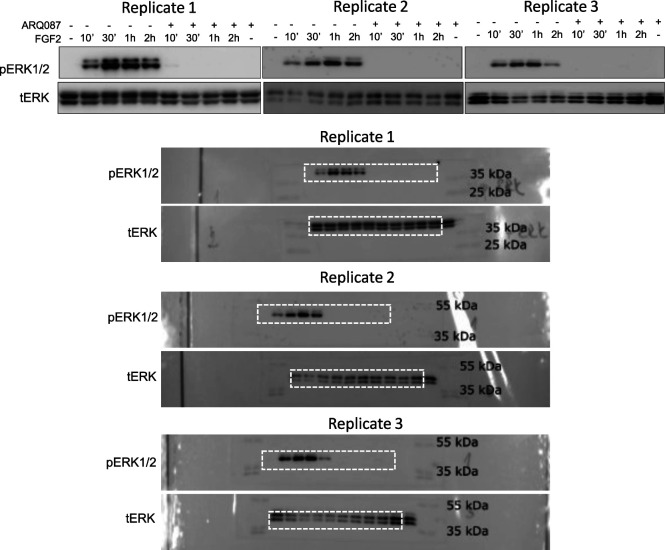
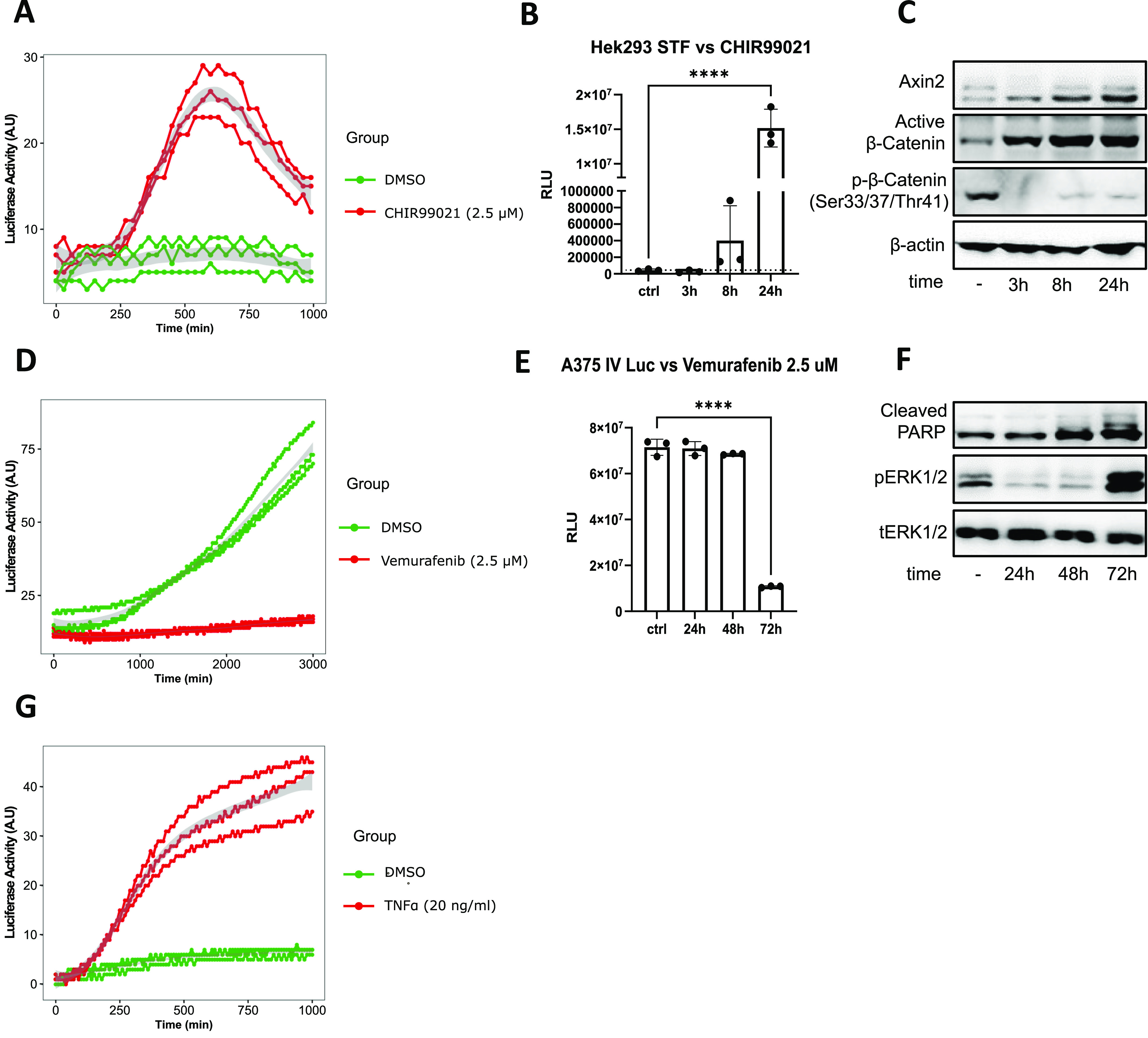
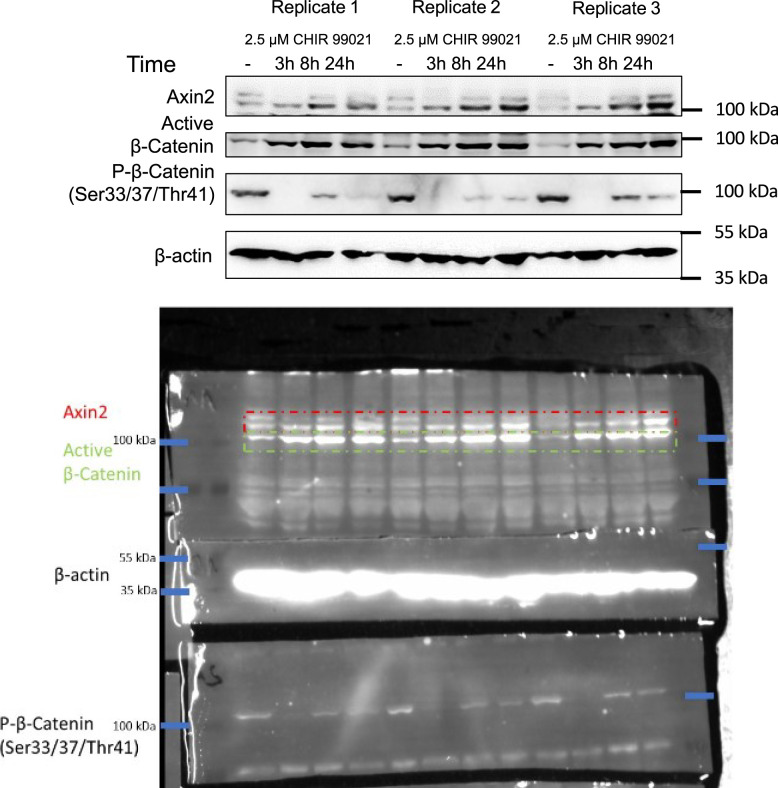
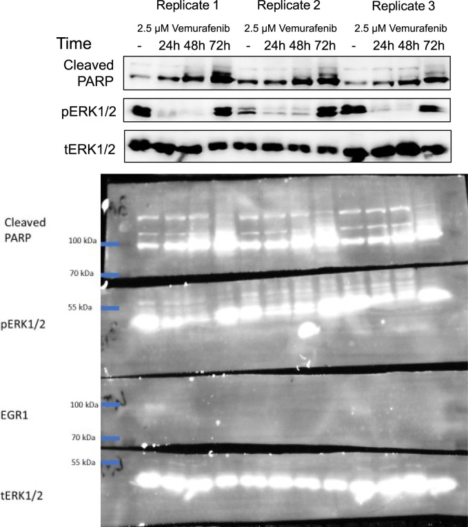
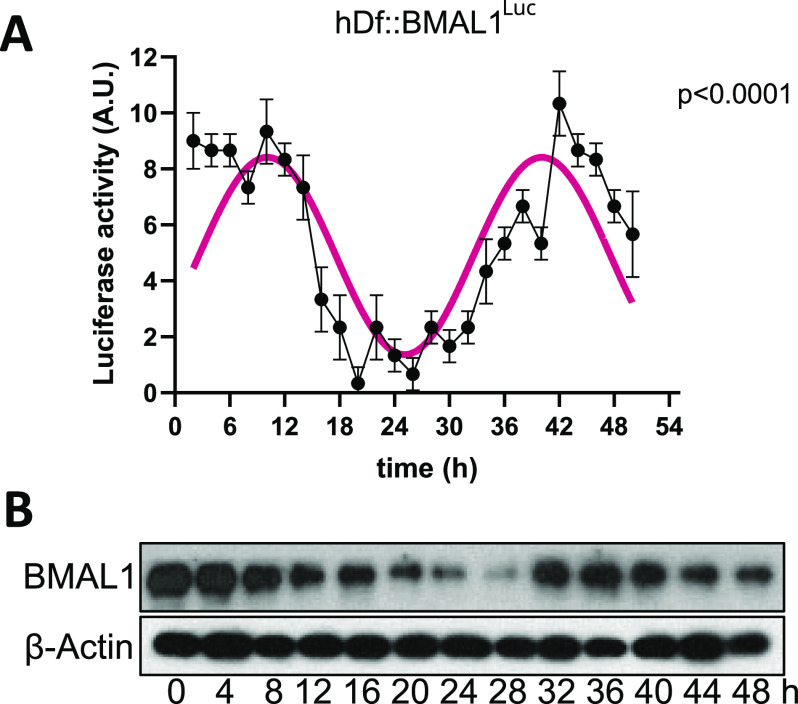
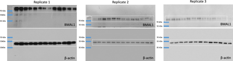
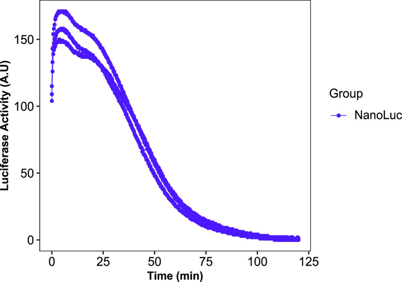
Luciferase reporter assays represent a simple and sensitive experimental system in cell and molecular biology to study multiple biological processes. However, the application of these assays is often limited by the costs of conventional luminometer instruments and the versatility of their use in different experimental conditions. Therefore, we aimed to develop a small, affordable luminometer allowing continuous measurement of luciferase activity, designed for inclusion into various kinds of tissue culture incubators. Here, we introduce LuminoCell-an open-source platform for the construction of an affordable, sensitive, and portable luminometer capable of real-time monitoring in-cell luciferase activity. The LuminoCell costs $40, requires less than 1 h to assemble, and it is capable of performing real-time sensitive detection of both magnitude and duration of the activity of major signalling pathways in cell cultures, including receptor tyrosine kinases (EGF and FGF), WNT/β-catenin, and NF-κB. In addition, we show that the LuminoCell is suitable to be used in cytotoxicity assays as well as for monitoring periodic circadian gene expression.


Conflict of interest statement

The authors declare that they have no conflict of interest.

Figures

Figure 1.
Figure 1.. The LuminoCell description.
(A) General wiring diagram. (B) The LuminoCell with lid (top view). (C) Cross section of the LuminoCell. green–lid, red–middle part, blue–bottom part.
Figure S1.
Figure S1.. Wiring diagram of the LuminoCell device.
Figure S2.
Figure S2.. Cross section of the LuminoCell.
Dimensions are given in millimetres.
Figure 2.
Figure 2.. Transactivation of pKrox24Luc by fibroblast growth factor receptor and epidermal growth factor receptor signalling.
(A) Luciferase activity upon addition of luciferin and recombinant FGF2 (arrows) to RCS::pKrox24Luc cells. fD, background (dark) frequency. Integration time for pulse count was 5 min, if not stated otherwise. (A, B) Normalized data from (A), with subtracted background. (C) FGF2-mediated pKrox24Luc transactivation in different cell densities. (D) 3,125 RCS::pKrox24Luc cells/cm2 were stimulated with 20 ng/ml FGF2, and the luciferase activity was measured using a 5 or 20 min integration time. (E) Luciferase activity in the presence of 20 ng/ml FGF2 and different concentrations of fibroblast growth factor receptor signalling inhibitor ARQ087 in RCS::pKrox24Luc cells. (F) Western blot analysis of ERK phosphorylation (p) in RCS::pKrox24Luc cells treated with 500 nM ARQ087 and 20 ng/ml FGF2. Total levels of ERK (t) serve as a loading control. Uncropped images are shown in Fig S3. (G) pKrox24Luc transactivation mediated by EGF in 293T::pKrox24Luc cells. Integration time 10 min. Data from three replicates are shown.
Figure S3.
Figure S3.. Western blot analysis of ERK phosphorylation (p) in RCS::pKrox24Luc cells treated with 500 nM ARQ087 and 20 ng/ml FGF2.
Total levels of ERK (t) represent a loading control. Dashed rectangle represents cropped area.
Figure 3.
Figure 3.. Testing the ability of the LuminoCell to measure luciferase activity of Wnt, TNFα pathways reporters, and for cytotoxicity testing.
(A) Assessment of canonical Wnt signalling activity in Super-Top-Flash (STF) cells in the presence of CHIR99021 or DMSO as a control. Luciferase activity measured using the LuminoCell (integration time 30 min). Data from three replicates are shown. (B) End-point analysis of canonical Wnt signalling activity in STF cells in the presence of CHIR99021 or DMSO as a control (ctrl). Luciferase activity measured using bench-top luminometer (Hidex Bioscan). (C) Western blot analysis of β-catenin and Axin2 expression in STF cells, β-actin represents a loading control. The data show representative blots of three independent experiments. Uncropped images are shown in Fig S4. (D) Cytotoxicity testing using A375-IVLuc cells. Cells were cultured in the presence of vemurafenib or DMSO as a control. Luciferase activity measured using the LuminoCell (integration time 20 min). Data from three replicates are shown. (E) End-point analysis of cytotoxicity using A375-IVLuc cells. Cells were cultured in the presence of vemurafenib or DMSO as a control. Luciferase activity measured using bench-top luminometer (Hidex Bioscan). (F) Western blot analysis of PARP cleavage and ERK (p) phosphorylation. Total (t) ERK represents a loading control. The data show representative blots of three independent experiments. Uncropped images are shown in Fig S5. (G) Assessment of NF-κB activity in the presence of TNFα using 293T::NF-κBLuc cells. Luciferase activity measured using the LuminoCell (integration time 10 min). Data from three replicates are shown.
Figure S4.
Figure S4.. Western blot analysis of β-catenin and Axin2 expression in Super-Top-Flash cells, β-actin represents a loading control.
Dashed rectangle represents cropped area.
Figure S5.
Figure S5.. Western blot analysis of PARP cleavage and ERK (p) phosphorylation.
Total (t) ERK represents a loading control. Dashed rectangle represents cropped area.
Figure 4.
Figure 4.. Testing the ability of the LuminoCell to quantify the activity of circadian BMAL1 luciferase reporter.
(A) Assessment of BMAL1 expression using luciferase reporter cell line hDf::BMAL1Luc upon serum shock (integration time 2 h). Error bars show mean ± sd from three replicates. (B) Expression of BMAL1 upon serum shock, as demonstrated using Western blot. β-actin represents loading control. The data show representative blots of three independent experiments. Uncropped images are shown in Fig S6.
Figure S6.
Figure S6.. Western blot analysis of BMAL1 expression in hDF upon serum shock treatment, β-actin represents a loading control.
Dashed rectangle represents cropped area.
Figure 5.
Figure 5.. Monitoring NanoLuc activity in ARPE-19 cells expressing NanoLuc (integration time 20 min).
Data from three replicates are shown.

References

    1. Balek L, Gudernova I, Vesela I, Hampl M, Oralova V, Bosakova MK, Varecha M, Nemec P, Hall T, Abbadessa G, et al. (2017) ARQ 087 inhibits FGFR signaling and rescues aberrant cell proliferation and differentiation in experimental models of craniosynostoses and chondrodysplasias caused by activating mutations in FGFR1, FGFR2 and FGFR3. Bone 105: 57–66. 10.1016/j.bone.2017.08.016 - DOI - PubMed
    1. Balsalobre A, Damiola F, Schibler U (1998) A serum shock induces circadian gene expression in mammalian tissue culture cells. Cell 93: 929–937. 10.1016/s0092-8674(00)81199-x - DOI - PubMed
    1. Bennett CN, Ross SE, Longo KA, Bajnok L, Hemati N, Johnson KW, Harrison SD, MacDougald OA (2002) Regulation of Wnt signaling during adipogenesis. J Biol Chem 277: 30998–31004. 10.1074/jbc.M204527200 - DOI - PubMed
    1. Brown SA, Fleury-Olela F, Nagoshi E, Hauser C, Juge C, Meier CA, Chicheportiche R, Dayer JM, Albrecht U, Schibler U (2005) The period length of fibroblast circadian gene expression varies widely among human individuals. PLoS Biol 3: e338. 10.1371/journal.pbio.0030338 - DOI - PMC - PubMed
    1. Chapman PB, Hauschild A, Robert C, Haanen JB, Ascierto P, Larkin J, Dummer R, Garbe C, Testori A, Maio M, et al. (2011) Improved survival with vemurafenib in melanoma with BRAF V600E mutation. N Engl J Med 364: 2507–2516. 10.1056/NEJMoa1103782 - DOI - PMC - PubMed

Publication types

LinkOut - more resources