. 2022 Apr 19;5(1):366.
doi: 10.1038/s42003-022-03299-8.

Evidence for postnatal neurogenesis in the human amygdala

Affiliations

Evidence for postnatal neurogenesis in the human amygdala

Sebastian S Roeder et al. Commun Biol. .

Abstract

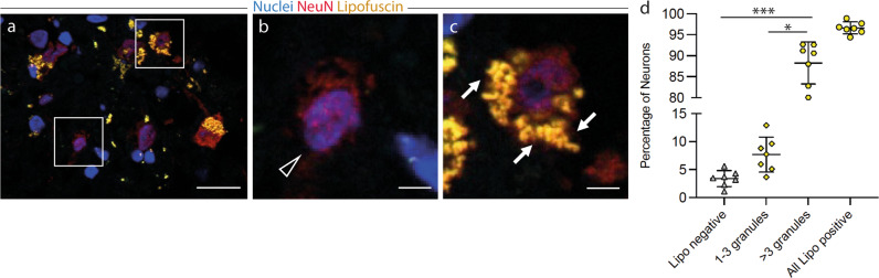
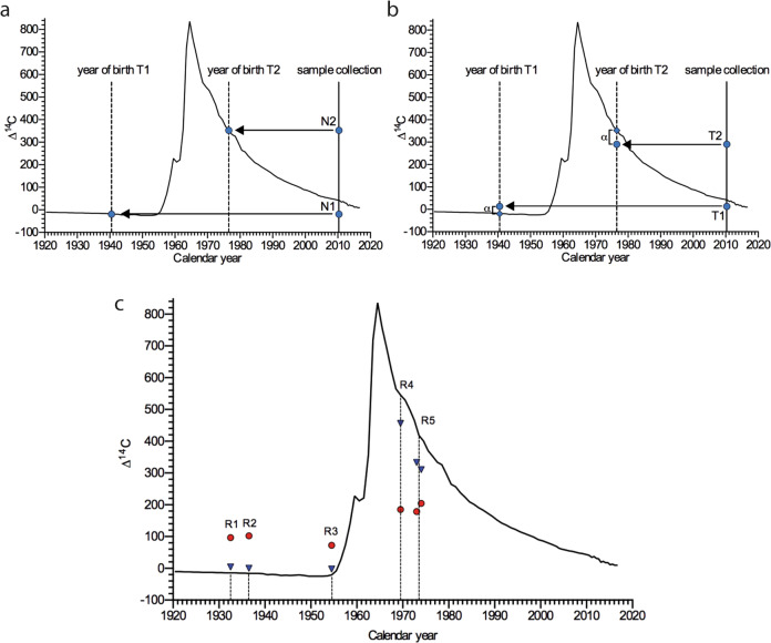
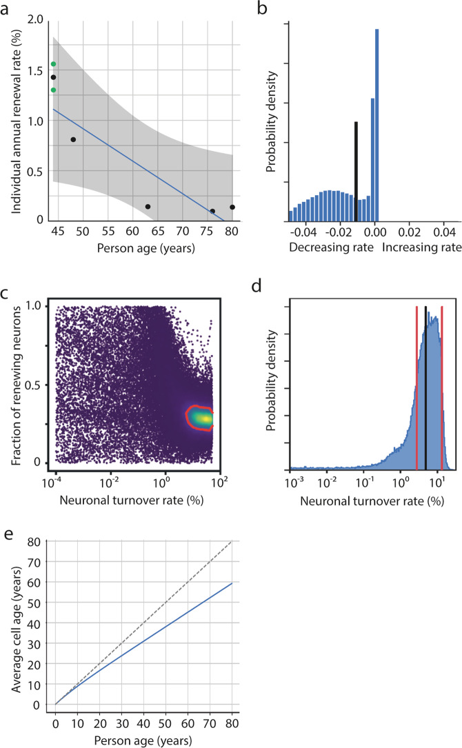
The human amygdala is involved in processing of memory, decision-making, and emotional responses. Previous studies suggested that the amygdala may represent a neurogenic niche in mammals. By combining two distinct methodological approaches, lipofuscin quantification and 14C-based retrospective birth dating of neurons, along with mathematical modelling, we here explored whether postnatal neurogenesis exists in the human amygdala. We investigated post-mortem samples of twelve neurologically healthy subjects. The average rate of lipofuscin-negative neurons was 3.4%, representing a substantial proportion of cells substantially younger than the individual. Mass spectrometry analysis of genomic 14C-concentrations in amygdala neurons compared with atmospheric 14C-levels provided evidence for postnatal neuronal exchange. Mathematical modelling identified a best-fitting scenario comprising of a quiescent and a renewing neuronal population with an overall renewal rate of >2.7% per year. In conclusion, we provide evidence for postnatal neurogenesis in the human amygdala with cell turnover rates comparable to the hippocampus.


Conflict of interest statement

The authors declare no competing interests.

Figures

Fig. 1
Fig. 1. Lipofuscin-negative neurons in the adult human amygdala indicate a neuronal cell age younger than the individual.
ad Lipofuscin positive and negative neurons in the human amygdala. Immunofluorescence staining of tissue sections of the adult human amygdala. Neurons were identified by NeuN-staining (red) and nuclei were labeled with 4’,6-diamidino-2-phenylindole (DAPI, blue). Presence of lipofuscin granules (yellow) was assessed by autofluorescence signal after excitation at 488 nm. a Boxed areas from the respective overview image, b showing lipofuscin-negative neurons (arrow heads), c and lipofuscin-positive neurons (arrows). d Quantification of lipofuscin granules per neuron (n = 7 patients, quantification of an average of 234 neurons/patient). Proportions of trichotomized lipofuscin levels for subjects L1-L7 (Supplemental Table 1) are presented as mean ± SEM; Kruskal–Wallis One Way Analysis of Variance on Ranks with post-hoc Tukey: *p < 0.05; ***p < 0.001. a Scale bar = 10 µm, b, c scale bar = 2 µm.
Fig. 2
Fig. 2. Genomic 14C-levels of neuronal and non-neuronal cells in the adult human amygdala provide evidence for post-natal neurogenesis.
a, b Interpretation of two hypothetical scenarios of differential cell turnover utilizing radiocarbon-based retrospective birth dating of cells. Atmospheric 14C concentrations of the last century are depicted by the black line. The vertical solid line represents the year of hypothetical sample collection. a In a no-cell-turnover-scenario the concentrations of genomic 14C in the year of sample collection of two individuals (N1, born 1940 and N2, born 1976) are identical to the atmospheric 14C levels in the years of birth of the individuals. The vertical dashed line represents the year of birth of the individuals and in this scenario meets the atmospheric 14C curve at the exact 14C concentration found at sample collection. b In a cell-turnover scenario the concentrations of genomic 14C levels in the year of sample collection of two individuals (T1, born 1940 and T2, born 1976) differ (alpha) from the atmospheric 14C levels in the years of birth of the individuals (vertical dashed line). In this turnover scenario the 14C levels of individuals born before the 14C peak are higher than the atmospheric 14C curve in the year of birth, whereas the 14C levels of individuals born after the 14C peak are below the atmospheric 14C curve in the year of birth of individuals. c 14C concentrations determined by accelerator mass spectrometry of twelve DNA samples derived from the amygdalae of five different, neurologically healthy subjects (R1-R5; bilateral amygdala of patient R5). Individual values are plotted at the time of birth of the respective subject (vertical dashed line). 14C levels of non-neuronal cells (red) and neuronal cells (blue) differ from the atmospheric 14C levels at the time of birth of the individuals, indicating post-natal neurogenesis.
Fig. 3
Fig. 3. Dynamics of neuronal cell turnover in the human amygdala.
Mathematical modelling of different neuronal cell turnover scenarios. a Scenario A assumes a constant turnover rate of neurons in the human amygdala over the lifetime of the individual. Fitting all samples to the model results in a median turnover rate of 0.2%/year [0.02;1.35]. However, separate fits for each sample revealed a negative correlation between age and individual turnover rate (r = −0.9, p = 0.083) with an annual decline in turnover of −0.03%/year. Green dots are amygdala measurements from both hemispheres from the same individual. Mean of these two dots are shown as black dot in between. b Scenario LIN, assuming a linear decline in cell turnover with age, revealed a median annual change in turnover rate (black vertical line) of −1 %/year [−4;0.08]. c Markov-Chain-Monte Carlo sampling for neuronal turnover for scenario 2POP, which assumes a quiescent and a renewing neuronal population, showing a two-dimensional marginal posterior distribution for turnover rate and fraction of renewing cells; blue: low probability, yellow: high probability, 1-sigma confidence region is framed by a red line. d Marginal posterior distribution for the estimated turnover rate calculated to the entire neuronal population, which is the product of the renewing rate of the renewing fraction, and the fraction of renewing cells, based on scenario POP2. Vertical red lines indicate lower and upper estimates (one sigma) of neuronal turnover. Black vertical line indicates median of turnover. e Age of amygdaloid neurons in relation to subject age based on the lower turnover limit of scenario 2POP. The bisecting (dashed) line represents a no-turnover-scenario in which every cell is as old as the individual, the blue line progressively deflects from the bisector with increasing subject age, reflecting lifelong post-natal neuronal cell turnover.

References

    1. Oppenheim RW. Adult hippocampal neurogenesis in mammals (and humans): the death of a central dogma in neuroscience and its replacement by a new dogma. Dev. Neurobiol. 2019;79:268–280. doi: 10.1002/dneu.22674. - DOI - PubMed
    1. Altman J, Das GD. Autoradiographic and histological evidence of postnatal hippocampal neurogenesis in rats. J. Comp. Neurol. 1965;124:319–335. doi: 10.1002/cne.901240303. - DOI - PubMed
    1. Gould E, Reeves AJ, Graziano MS, Gross CG. Neurogenesis in the neocortex of adult primates. Science. 1999;286:548–552. doi: 10.1126/science.286.5439.548. - DOI - PubMed
    1. Eriksson PS, et al. Neurogenesis in the adult human hippocampus. Nat. Med. 1998;4:1313–1317. doi: 10.1038/3305. - DOI - PubMed
    1. Spalding KL, et al. Dynamics of hippocampal neurogenesis in adult humans. Cell. 2013;153:1219–1227. doi: 10.1016/j.cell.2013.05.002. - DOI - PMC - PubMed

Publication types