Review
. 2022 Jun;23(6):323-341.
doi: 10.1038/s41583-022-00576-7. Epub 2022 Apr 19.

Genomics, convergent neuroscience and progress in understanding autism spectrum disorder

Affiliations
Review

Genomics, convergent neuroscience and progress in understanding autism spectrum disorder

Helen Rankin Willsey et al. Nat Rev Neurosci. 2022 Jun.

Abstract

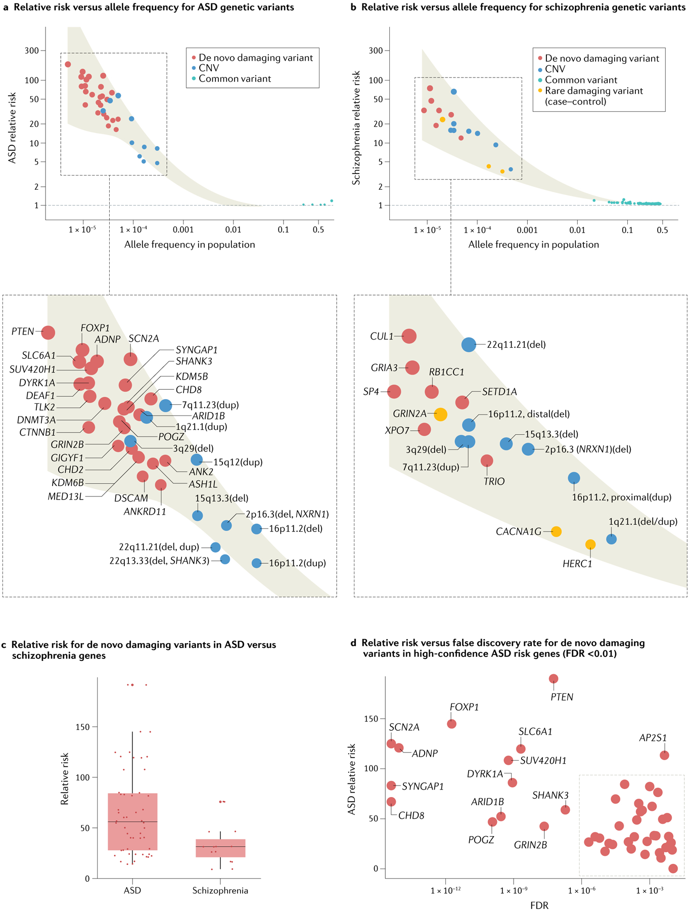
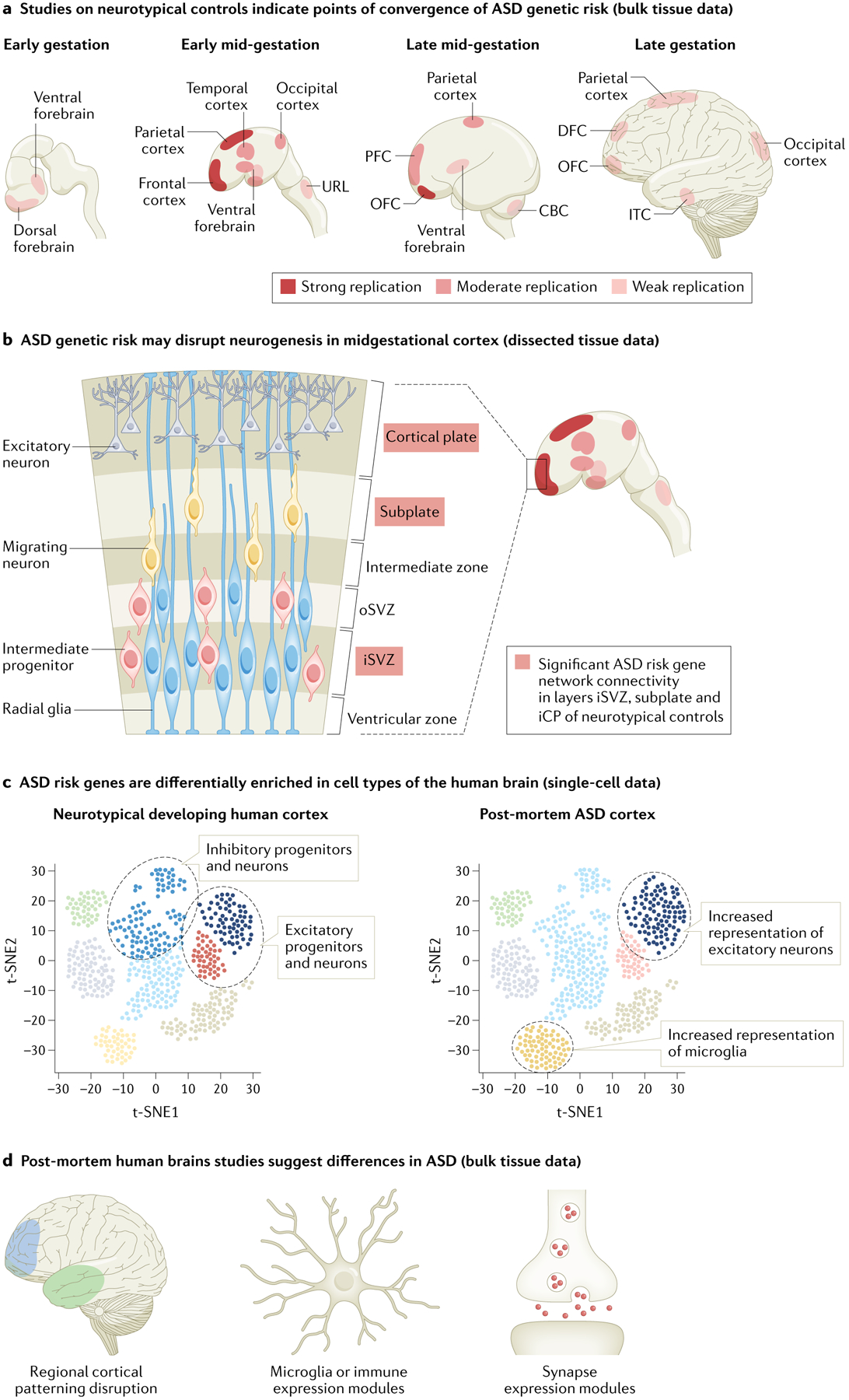
More than a hundred genes have been identified that, when disrupted, impart large risk for autism spectrum disorder (ASD). Current knowledge about the encoded proteins - although incomplete - points to a very wide range of developmentally dynamic and diverse biological processes. Moreover, the core symptoms of ASD involve distinctly human characteristics, presenting challenges to interpreting evolutionarily distant model systems. Indeed, despite a decade of striking progress in gene discovery, an actionable understanding of pathobiology remains elusive. Increasingly, convergent neuroscience approaches have been recognized as an important complement to traditional uses of genetics to illuminate the biology of human disorders. These methods seek to identify intersection among molecular-level, cellular-level and circuit-level functions across multiple risk genes and have highlighted developing excitatory neurons in the human mid-gestational prefrontal cortex as an important pathobiological nexus in ASD. In addition, neurogenesis, chromatin modification and synaptic function have emerged as key potential mediators of genetic vulnerability. The continued expansion of foundational 'omics' data sets, the application of higher-throughput model systems and incorporating developmental trajectories and sex differences into future analyses will refine and extend these results. Ultimately, a systems-level understanding of ASD genetic risk holds promise for clarifying pathobiology and advancing therapeutics.


Conflict of interest statement

Competing interests

The authors declare no competing interests.

Figures

Fig. 1 |
Fig. 1 |. Relationship between effect size and allele frequency for loci discovered in autism spectrum disorder or schizophrenia.
a,b | Systematic studies of rare and common variants have identified risk loci for autism spectrum disorder (ASD) and for schizophrenia, but the trajectory of discovery differs between these conditions. In ASD, the majority of loci discovered to date are genes and have been identified by exome-wide sequencing studies of rare, generally de novo, coding variants (P < 2.5 × 10⁻⁶; red dots; for ASD, 26 genes; for schizophrenia, 10 genes), whereas in schizophrenia the majority are single-nucleotide polymorphisms (SNPs) and have been discovered based on genome-wide association studies (GWAS) of common variants (P < 5 × 10⁻⁸; teal dots; for ASD, 5 SNPs; for schizophrenia, 270 SNPs, of which only the 132 SNPs with relative risk >1 are shown). None of these genes (red) or SNPs (teal) overlap between ASD and schizophrenia. Both disorders have a similar number of loci associated based on rare, generally de novo, copy number variants (CNVs; blue dots; for ASD, permutation-based false discovery rate (FDR) ≤ 0.02, 10 loci; for schizophrenia, Benjamini–Hochberg FDR ≤ 0.02, 8 loci). In contrast to rare coding variants and common variants, most of the top CNVs overlap between ASD and schizophrenia. In general, rare variants carry substantially higher relative risks than common variants. c | De novo damaging variants in the top 26 ASD risk genes tend to carry higher relative risk than de novo damaging variants within the top 7 schizophrenia risk genes (de novo damaging variants have been identified in only 7 out of the 10 schizophrenia risk genes), underscoring the particularly large contribution of rare variants to ASD. d | Utilizing a conservative FDR threshold instead of an exome-wide significant P value (FDR ≤ 0.01 versus P < 2.5 × 10⁻⁶) highlights 47 high-confidence ASD risk genes, with varying relative risks. In general, the most strongly associated genes carry the largest relative risks. See Supplementary Table 1 for a complete list of gene names, FDRs and effect sizes; see REF. for a broader list of ASD risk genes as determined by FDR. Note that we defined relative risk as the ratio of the frequency of a given variant in cases versus unaffected controls. Dot size is proportional to relative risk. Shaded area represents 95% confidence interval of the locally weighted least squares regression. Location on x axis in parts a,b is based on frequency in unaffected controls. As there are no associations in ASD for alleles with frequencies between 0.001 and 0.1, no curve is shown in this interval in part a. Damaging variants consist of protein-truncating variants (frameshift, canonical splice-acceptor, canonical splice-donor and nonsense variants) and missense 3 (Mis3) variants (those predicted to be probably damaging by PolyPhen-2 (REF.)). To estimate the per gene relative risk for de novo damaging variants, we defined the frequency in cases as the total number of de novo damaging variants observed in a given gene, and the frequency in controls based on estimates of the number of mutations expected per generation. All 26 genes identified in ASD have multiple de novo damaging variants in probands. However, only seven of ten genes identified in schizophrenia have de novo variants (the rest were identified by rare damaging variants in case–control data only). Therefore, we estimated relative risk for de novo damaging variants when possible (red dots) and for rare damaging variants when necessary (orange dots). To estimate relative risk for rare damaging variants (case–control, schizophrenia only), we defined the frequency of each allele in cases based on reported frequency for schizophrenia and the frequency in controls based on the frequency observed in the non-psychiatric subset of the gnomAD v2 data set. To estimate relative risk for rare CNVs, we defined the frequency in cases based on reported frequencies for ASD and for schizophrenia and the frequency in controls based on frequency observed in the Database of Genomic Variants (DGV) (ASD) or directly from controls (schizophrenia). We estimated the 3q29(del) frequency in schizophrenia as 0.00005 because no 3q29(dels) were observed in 20,227 controls. The 15q13.3(del) is referred to as 15q13.2–13.3 in REF. and we estimated its frequency as 0.000092 based on structural variants in gnomAD because of inconsistent frequencies reported in smaller studies in DGV. We estimated relative risk for common variants based on reported odds ratios for ASD or for schizophrenia. Part b adapted from REF., CC BY 4.0 (https://creativecommons.org/licenses/by/4.0/).
Fig. 2 |
Fig. 2 |. Emerging patterns of ASD convergence identified from gene expression data.
Numerous studies investigating human gene expression data sets for convergence related to autism spectrum disorder (ASD) risk have been conducted, spanning so-called ‘neurotypical’ (control) brain samples (parts ac) as well as post-mortem brain samples from individuals with ASD (parts c,d). These data sets encompass bulk tissue from major regions of the brain (parts a,d), bulk tissue from more finely dissected regions of the developing cortex (part b) and single cells from the cortex (part c). Neurotypical data tend to include prenatal and postnatal samples across the entire spectrum of human brain development whereas patient-derived data include postnatal samples only (ASD diagnosis requires behavioural assessment). Generally, approaches utilizing neurotypical expression data seek to identify convergence by searching for ‘enrichment’ of ASD risk genes in particular developmental epochs, brain regions and/or cell types. In this case, enrichment is indicated by the ‘strength’ of co-expression of ASD risk genes or by ‘high’ or ‘specific’ expression of ASD risk genes. By contrast, approaches utilizing patient expression data generally aim to identify convergence by characterizing systematic differences between cases and matched controls (that is, genes consistently differentially expressed in brain tissue from individuals with ASD). Although data and approaches differ substantially across studies, the developing frontal cortex and excitatory neurons are recurring points of enrichment. a | Studies investigating spatiotemporal convergence of ASD genetic risk in gene expression data from bulk samples of neurotypical brains have consistently highlighted mid-gestational frontal and parietal cortices,,,,,,,. We summarize the extent of replication by color intensity, with the strongest replication indicated by dark red and the weakest by light red. These studies varied widely in granularity of developmental periods and brain regions studied, and therefore we considered studies that examined two or fewer regional (for example, grouping all cortical regions, studying only one brain region) or temporal (for example, prenatal versus postnatal) contexts to provide ‘limited’ evidence. Strong replication denotes a significant finding in at least three ‘non-limited’ studies, moderate replication indicates a significant finding in two non-limited studies and weak replication corresponds to a significant finding in one non-limited study and at least one limited study. We define early gestation as 8–10 post-conception weeks (PCW), early mid-gestation as 10–16 PCW, late mid-gestation as 16–24 PCW and late gestation as 24–38 PCW. b | Based on findings summarized in part a, several studies have examined laminar convergence of ASD genetic risk in neurotypical early mid-gestational frontal cortex. An initial study assessing preservation of spatiotemporal co-expression networks by layer implicated the inner cortical plate whereas a contemporaneous study assessing specificity of expression of ASD gene lists by layer indicated a relative lack of layer-specific enrichment. A later study incorporating orthogonal gene interaction data and assessing ASD gene network connectivity by layer replicated the inner cortical plate finding and newly implicated the inner subventricular zone and subplate. c | Analyses of single-cell gene expression data from neurotypical controls and individuals with ASD have consistently highlighted excitatory neurons. In neurotypical mid-gestational cortex, ASD risk genes tend to be highly and/or specifically expressed in developing excitatory and inhibitory neurons (progenitors and maturing neurons),,,,. Analyses of single-cell gene expression data from (postnatal) cortex of individuals with ASD have similarly identified consistent differences between cases and controls in excitatory neurons, as well as potential differences in microglia and oligodendrocytes,. d | Analyses of bulk samples of brain tissue from individuals with ASD compared with those from matched neurotypical controls support the major findings from parts ac. These studies implicate frontal cortex, excitatory and inhibitory neurons, and microglia, and additionally highlight differences in genes associated with astrocytes and in expression modules related to synaptic function and inflammation–,. CBC, cerebellar cortex; DFC, dorsal frontal cortex; iCP, inner cortical plate; iSVZ, inner subventricular zone; ITC, inferior temporal cortex; OFC, orbital frontal cortex; oSVZ, outer subventricular zone; PFC, prefrontal cortex; URL, upper rhombic lip. Some of the brain images in parts a,b and d are adapted with permission from REF.

References

    1. Lord C et al. Autism spectrum disorder. Nat. Rev. Dis. Prim 6, 5 (2020). - PMC - PubMed
    1. Díaz-Caneja CM et al. A white paper on a neurodevelopmental framework for drug discovery in autism and other neurodevelopmental disorders. Eur. Neuropsychopharmacol 48, 49–88 (2021). - PubMed
    1. Folstein S & Rutter M Infantile autism: a genetic study of 21 twin pairs. J. Child. Psychol. Psychiatry 18, 297–321 (1977). - PubMed
    1. Gaugler T et al. Most genetic risk for autism resides with common variation. Nat. Genet 46, 881–885 (2014). - PMC - PubMed
    1. Klei L et al. Common genetic variants, acting additively, are a major source of risk for autism. Mol. Autism 3, 9 (2012). - PMC - PubMed

Publication types