. 2022 Feb 18;13(14):3965-3976.
doi: 10.1039/d1sc06182h. eCollection 2022 Apr 6.

Template directed synthesis of antibody Fc conjugates with concomitant ligand release

Affiliations

Template directed synthesis of antibody Fc conjugates with concomitant ligand release

Viktoriia Postupalenko et al. Chem Sci. .

Abstract

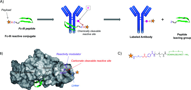
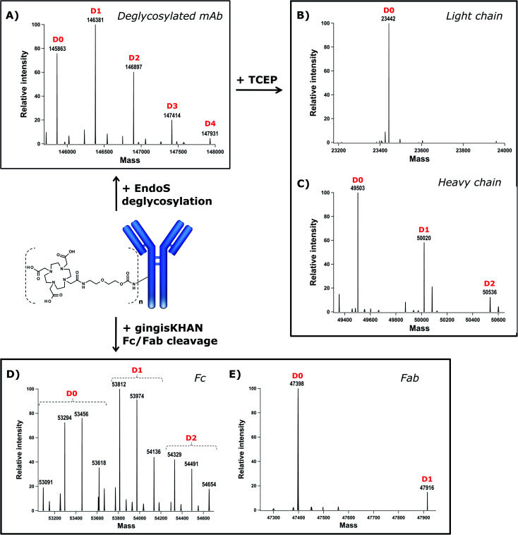
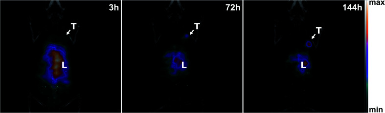
Antibodies are an attractive therapeutic modality for cancer treatment as they allow the increase of the treatment response rate and avoid the severe side effects of chemotherapy. Notwithstanding the strong benefit of antibodies, the efficacy of anti-cancer antibodies can dramatically vary among patients and ultimately result in no response to the treatment. Here, we have developed a novel means to regioselectively label the Fc domain of any therapeutic antibody with a radionuclide chelator in a single step chemistry, with the aim to study by SPECT/CT imaging if the radiolabeled antibody is capable of targeting cancer cells in vivo. A Fc-III peptide was used as bait to bring a carbonate electrophilic site linked to a metal chelator and to a carboxyphenyl leaving group in close proximity with an antibody Fc nucleophile amino acid (K317), thereby triggering the covalent linkage of the chelator to the antibody lysine, with the concomitant release of the carboxyphenyl Fc-III ligand. Using CHX-A''-DTPA, we radiolabeled trastuzumab with indium-111 and showed in biodistribution and imaging experiments that the antibody accumulated successfully in the SK-OV-3 xenograft tumour implanted in mice. We found that our methodology leads to homogeneous conjugation of CHX-A''-DTPA to the antibody, and confirmed that the Fc domain can be selectively labeled at K317, with a minor level of unspecific labeling on the Fab domain. The present method can be developed as a clinical diagnostic tool to predict the success of the therapy. Furthermore, our Fc-III one step chemistry concept paves the way to a broad array of other applications in antibody bioengineering.


Conflict of interest statement

There are no conflicts to declare. V. P, L. M, F. L, P. G, J.-M. S, and O. N are coinventors of application WO2021110860 A1 filed on 3 December 2019, entitled “Reactive conjugates”.

Figures

Fig. 1
Fig. 1. (A) Schematic representation of the one-step antibody labeling approach. The payload is attached to the Fc-III peptide via a chemically reactive site “X–Z”. X in red refers to the electrophilic site, and Z in violet refers to the leaving group. Upon binding of the Fc-III reactive conjugate to IgG-Fc, the antibody lysine is poised to attack the electrophilic center of the chemically reactive site X, and the peptide vector is detached from the payload thanks to the engineered leaving group Z. This strategy allows subsequent wash out of the ligand without any additional steps. (B) Representation of the binding of Fc-III to the Fc domain of IgG based on the X-ray structure. The surface of IgG-Fc is shaded grey, and Fc-III is shown in a green ribbon structure. The antibody K317 is shown with a pink sphere. The PEG spacer is linked to a carbonate reactive site via a reactivity modulator that jointly ensures high reactivity in the presence of lysine residues and adequate stability of the reactive site towards hydrolysis. The payload (yellow star), typically a fluorescent label (fluorescein), a radiolabel, or a chelator (DOTA, DTPA, and DFO), is chemically bound to the other side of the carbonate reactive group via a short linker. (C) Chemical structure of the payload-PEGn-Fc-III reactive conjugate using the same color codes as in A and B.
Scheme 1
Scheme 1. Synthesis of the FITC-PEG20-Fc-III reactive conjugate: (a) HATU, DIEA, DMF, and then Fc-III peptide; (b) 20% piperidine, DMF; (c) compound 4, Et3N in ACN at 40 °C; (d) compound 6, DMAP in DCM at 25 °C; (e) TFA/DCM (1/3); (f) FITC, DIEA in ACN/DMF 1/1 at 25 °C; (g) HATU, DIEA, DMF, and NH2-PEG20-Fc-III peptide.
Fig. 2
Fig. 2. (A) Binding isotherm of Fc-III-FAM to trastuzumab measured by fluorescence anisotropy. The trastuzumab concentration is gradually increased while the concentration of the Fc-III-FAM labeled peptide was kept constant (5 nM). The fluorescence polarization signal of the trastuzumab/peptide bound complexes is reported on the y axis. The black solid line corresponds to the fit of the data using the Hill equation, yielding the half-maximal effective concentration (EC50). (B) Competition assays of Fc-III and PEG20-Fc-III for trastuzumab against Fc-III-FAM. Black solid (Fc-III) and red dashed lines (PEG20-Fc-III) are fittings of the data using the Hill equation yielding the half-maximal inhibitory concentration (IC50).
Fig. 3
Fig. 3. Optimization of the reaction conditions for trastuzumab conjugation with FITC-PEG20-Fc-III. (A and C) SDS gel electrophoresis of fluorescently labeled antibody obtained after increasing the incubation time (top panel) and peptide equivalents (bottom panel). Derivatized mAbs were run under the reducing conditions, and fluorescence was measured using the FluoroM bio-imaging system. The bands next to the 50 and 25 kDa marker correspond to Hc and Lc, respectively. (B and D) Quantification of the band fluorescence intensities of the SDS gels of panels A and C, respectively. The incubation time and peptide equivalents of each band of panels A (from right to left) and C correspond to the data points of panels B and D , respectively (Fig. S2†). The FITC-carbonate data (B and D) were extracted from the gels in Fig. S2.
Fig. 4
Fig. 4. Intact mass analysis of the trastuzumab-DOTA conjugate and its subunits by LC-FTMS. The deconvolved mass spectra of the intact deglycosylated sample (A) and deglycosylated TCEP reduced sample, yielding light (B) and heavy chains (C); GingisKHAN™ derived Fc (D) and Fab (E) subunits. The D0–D4 peaks correspond to the antibody or antibody fragment with different degrees of conjugation, where Dn is the number of attached DOTA molecules.
Fig. 5
Fig. 5. Effect of the spacer length on Fc/F(ab')2 selectivity. The spacer length calculated with the WLC model is plotted on the x-axis relative to the Fc/F(ab')2 selectivity ratio. The number of PEG units is color coded in the legend box according to its length.
Fig. 6
Fig. 6. A constant amount of the radioligand was incubated with an increasing concentration of SK-OV-3 (HER2-positive) and MDA-MB-231 (HER2-negative) cells. The specific cell binding is expressed as a function of the number of cells. A binding curve (dashed line) is fitted using nonlinear regression, and the immunoreactive fraction corresponding to the extrapolation to infinite antigen excess is calculated using Graphpad Prism 7 software. 81% of the immunoreactive fraction of [111In]In-DTPA-trastuzumab for SK-OV-3 showed a conserved affinity for HER2 after DTPA regiospecific conjugation and radiolabeling with [111In]indium. The immunoreactivity decreased to 15% with MDA-MB-231 (HER2-negative).
Fig. 7
Fig. 7. Coronal MIP fused SPECT and CT images at 3, 72, and 144 h post-injection of 18 MBq [111In]In-DTPA-trastuzumab in the same mouse bearing an SK-OV-3 tumour (HER2-positive). Uptake of [111In]In-DTPA-trastuzumab was observed in the tumour (T) and liver (L). The longer blood circulation of [111In]In-DTPA-trastuzumab leads to an increased specific tumour uptake over time.

References

    1. Sung H. Ferlay J. Siegel R. L. Laversanne M. Soerjomataram I. Jemal A. Bray F. Ca-Cancer J. Clin. 2021;71:209–249. doi: 10.3322/caac.21660. - DOI - PubMed
    1. Chabner B. A. Roberts Jr T. G. Nat. Rev. Cancer. 2005;5:65–72. doi: 10.1038/nrc1529. - DOI - PubMed
    1. Tsimberidou A. M. Fountzilas E. Nikanjam M. Kurzrock R. Cancer Treat. Rev. 2020;86:102019. doi: 10.1016/j.ctrv.2020.102019. - DOI - PMC - PubMed
    1. Scott A. M. Wolchok J. D. Old L. J. Nat. Rev. Cancer. 2012;12:278–287. doi: 10.1038/nrc3236. - DOI - PubMed
    1. Dietel M. Johrens K. Laffert M. V. Hummel M. Blaker H. Pfitzner B. M. Lehmann A. Denkert C. Darb-Esfahani S. Lenze D. Heppner F. L. Koch A. Sers C. Klauschen F. Anagnostopoulos I. Cancer Gene Ther. 2015;22:417–430. doi: 10.1038/cgt.2015.39. - DOI - PubMed