Case Reports
. 2022 Apr 15;10(4):e05737.
doi: 10.1002/ccr3.5737. eCollection 2022 Apr.

Differentiation of intracranial Rosai-Dorfman histiocytosis from meningioma using MR perfusion

Affiliations
Case Reports

Differentiation of intracranial Rosai-Dorfman histiocytosis from meningioma using MR perfusion

Hugo F Bueno et al. Clin Case Rep. .

Abstract

Intracranial Rosai-Dorfman disease may be indistinguishable from meningioma. This distinction is essential, as they are treated very differently. We present two cases where perfusion imaging helped make this distinction, allowing one to be treated successfully without craniotomy. Perfusion imaging may be a powerful adjunct in cases where RDD mimics meningioma.

Keywords: MR perfusion; Rosai‐Dorfman disease; histiocytosis; meningioma.


Conflict of interest statement

The authors have no personal, financial, or institutional disclosures in relation to this article.

Figures
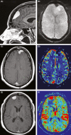
FIGURE 1
FIGURE 1
Case 1. 50‐year‐old man with history of RDD. (A) Sagittal postcontrast image showing a dural‐based enhancing extra‐axial mass with a dural tail. (B) Axial SWI demonstrates hypointense signal at the site of the lesion, suggesting mineralization, typically seen with meningiomas. (C) Axial postcontrast image again shows the enhancing mass which demonstrates (D) decreased relative cerebral blood volume (rCBV) compared to surrounding tissue. In contrast, (E) a typical meningioma demonstrates (F) markedly increased rCBV
FIGURE 2
FIGURE 2
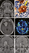
Case 2. (A) Coronal image through the paranasal sinuses demonstrates a mass filling the sinonasal cavity. (B) Pathologic specimen of the sinonasal mass with S‐100 immunostain demonstrates emperipolesis with intracytoplasmic lymphocytes, compatible with RDD. (C) Axial postcontrast image through the mass demonstrates avid enhancement, and (D) MR perfusion shows decreased rCBV. (E) Coronal postcontrast image through the frontal lobes demonstrates an avidly enhancing, dural‐based mass encasing the supraclinoid segment of the internal carotid artery. (F) Repeat imaging at 18 months demonstrates marked decrease in volume of the mass after treatment
FIGURE 3
FIGURE 3
Case 2 tumor volume and response to therapy over time. This graph demonstrates tumor volume as measured in cm3 and changes in time with red arrows noting points where therapeutic changes were initiated or made. The red block between weeks 40 and 44 denotes the time where radiation therapy was administered

References

    1. Foucar E, Rosai J, Dorfman R. Sinus histiocytosis with massive lymphadenopathy (Rosai‐Dorfman disease): review of the entity. Semin Diagn Pathol. 1990;7:19‐73. - PubMed
    1. Andriko J‐AW, Morrison A, Colegial CH, Davis BJ, Jones RV. Rosai‐Dorfman disease isolated to the central nervous system: a report of 11 cases. Mod Pathol. 2001;14:172‐178. - PubMed
    1. Zhu H, Qiu L‐H, Dou Y‐F, et al. Imaging characteristics of Rosai‐Dorfman disease in the central nervous system. Eur J Radiol. 2012;81:1265‐1272. - PubMed
    1. Rosai J, Dorfman RF. Sinus histiocytosis with massive lymphadenopathy. A newly recognized benign clinicopathological entity. Arch Pathol. 1969;87:63‐70. - PubMed
    1. Tatit RT, Raffa P, de Almeida Motta GC, et al. Rosai‐Dorfman disease mimicking images of meningiomas: two case reports and literature review. Surg Neurol Int. 2021;12:292. - PMC - PubMed

Publication types

LinkOut - more resources