. 2022 Apr 15;3(2):e125.
doi: 10.1002/mco2.125. eCollection 2022 Jun.

Regulation of XPO5 phosphorylation by PP2A in hepatocellular carcinoma

Affiliations

Regulation of XPO5 phosphorylation by PP2A in hepatocellular carcinoma

Jiao Li et al. MedComm (2020). .

Abstract

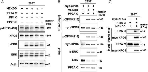
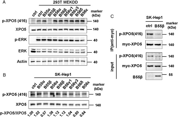
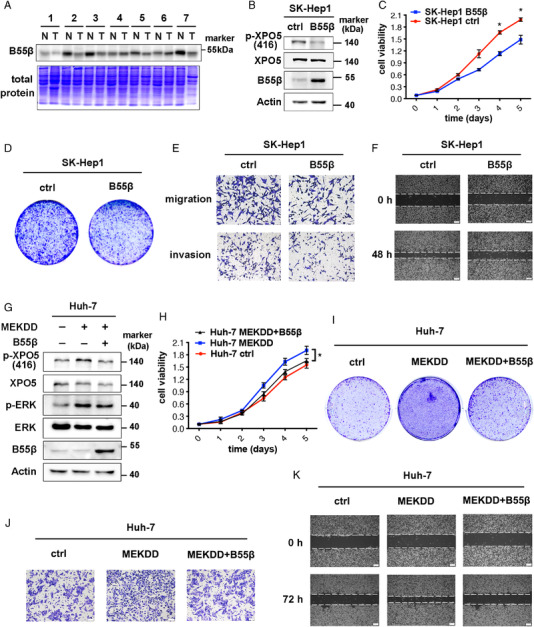
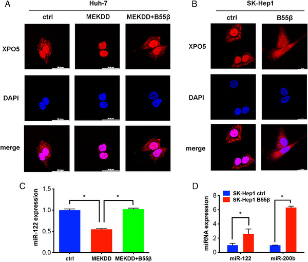
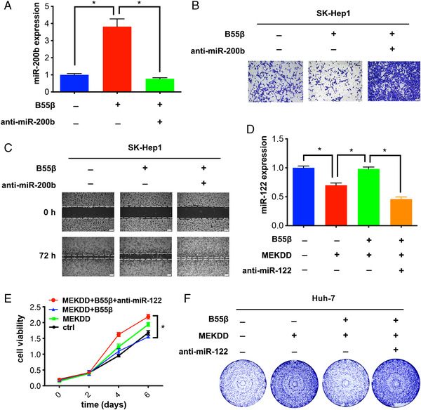
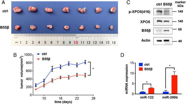
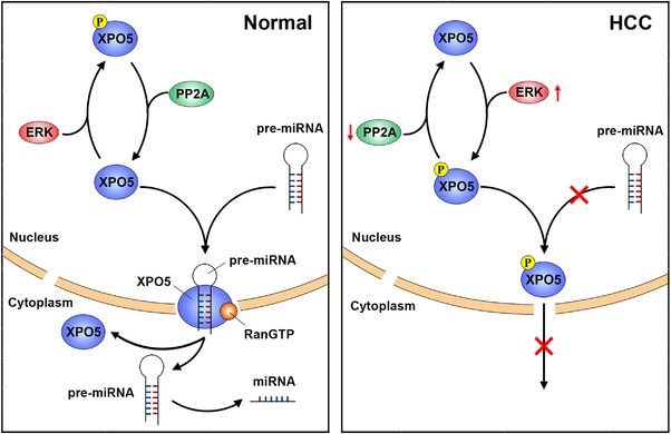
Exportin 5 (XPO5) is a shuttle protein that mediates precursor miRNA (pre-miRNA) export from the nucleus to the cytoplasm, an important step in miRNA maturation. We previously demonstrated that XPO5 was phosphorylated by ERK kinase and subsequently underwent conformation change by the peptidyl-prolyl isomerase Pin1, leading to the reduced miRNA expression in hepatocellular carcinoma (HCC). Protein phosphorylation modification serves as a reversible regulatory mechanism precisely governed by protein kinases and phosphatases. Here we identified that the phosphatase PP2A catalyzed XPO5 dephosphorylation. PP2A holoenzyme is a ternary complex composed of a catalytic subunit, a scaffold subunit, and a regulatory subunit that determines substrate specificity. In this study, we characterized the involvement of B55β subunit in XPO5 dephosphorylation that favored the distribution of XPO5 into the cytoplasm and promoted miRNA expression, leading to HCC inhibition in vitro and in vivo. Our study demonstrates the regulatory role of B55β-containing PP2A in miRNA expression and may shed light on HCC pathogenesis.

Keywords: PP2A; XPO5; hepatocellular carcinoma; microRNA.


Conflict of interest statement

The author Yong Peng is an associate editor of MedComm and not involved in the journal's review of, or decisions related to, this manuscript. The authors declare no conflict of interest.

Figures

FIGURE 1
FIGURE 1
PP2A catalyzes the dephosphorylation of XPO5. (A) HEK‐293T cells were transfected with plasmids expressing MEKDD and different catalytic subunits of serine/threonine phosphatases (PP1, PP2B, and PP2A). The phosphorylated and total XPO5/ERK proteins were detected via Western blot. (B) HEK‐293T cells were transfected with plasmids expressing MEKDD, myc‐XPO5 and C subunits of PP2A and myc‐XPO5 was immunoprecipitated from cell lysates. The immunoprecipitates were immunoblotted with the indicated antibodies. (C) The lysates from HEK‐293T cells co‐transfected with the indicated plasmids were subjected to immunoprecipitation via anti‐myc antibody. The enriched complexes were immunoblotted with the indicated antibodies. MEKDD: constitutively active MEK; p‐XPO5 (416): phosphorylated XPO5 at serine 416.
FIGURE 2
FIGURE 2
B55β is specifically engaged in XPO5 dephosphorylation. (A) HEK‐293T cells were transfected with plasmids expressing MEKDD and different regulatory subunits. The phosphorylated and total XPO5/ERK proteins were detected via Western blot. (B) SK‐Hep1 cells were transfected with plasmids expressing different regulatory subunits. The phosphorylated and total XPO5 proteins were detected via Western blot and the protein bands were quantified by ImageJ software. Comparison of the intensity of p‐XPO5 bands (using total XPO5 as loading control) was indicated as fold change relative to ctrl set. (C) SK‐Hep1 cells were co‐transfected with the indicated plasmids and myc‐XPO5 was immunoprecipitated from cell lysates. The immunoprecipitates were immunoblotted with the indicated antibodies.
FIGURE 3
FIGURE 3
B55β inhibits growth, migration and invasion abilities of HCC cells. (A) The protein lysates of human HCC and adjacent normal tissues were subjected to SDS‐PAGE and then stained with coomassie blue or immunoblotted with anti‐B55β antibody. (B) B55β level was determined by Western blot in SK‐Hep1 stable cell lines with or without B55β overexpression. (C–F) MTT assays (C), cloning formation (D), transwell migration and invasion assays (E), and wound healing (F) for SK‐Hep1 B55β overexpression and ctrl cells. (G) B55β level was detected by Western blot in Huh‐7 stable cell lines with or without MEKDD/B55β overexpression. (H‐K) MTT assays (H), cloning formation (I), transwell migration assays (J), and wound healing (K) for Huh‐7 stable cell lines with or without MEKDD/B55β overexpression. Data are shown as the means ± SD. *p < 0.05.
FIGURE 4
FIGURE 4
B55β regulates XPO5 distribution and miRNA expression. (A) Immunofluorescence of subcellular localization of XPO5 (Red) in Huh‐7 cells transfected with the indicated plasmids. (B) Immunofluorescence of subcellular localization XPO5 (Red) in SK‐Hep1 Ctrl and B55β overexpression cells. (C) The miR‐122 expression in Huh‐7 cells transfected with the indicated plasmids was determined by qRT‐PCR. (D) The expression of miR‐122 and miR‐200b in SK‐Hep1 ctrl and B55β overexpression cells was examined by qRT‐PCR. Data are shown as the means ± SD. *p < 0.05, **p < 0.01.
FIGURE 5
FIGURE 5
B55β‐induced HCC inhibition is partially attributed to the regulation of miRNA expression. (A) SK‐Hep1 ctrl and B55β overexpression cells were transfected with or without anti‐miR‐200b, followed by the examination of miR‐200b expression via qRT‐PCR. (B) Transwell migration assays and (C) wound healing assays for SK‐Hep1 stable cell lines transfected with or anti‐miR‐200b. (D) miR‐122 expression was detected by qRT‐PCR in Huh‐7 cells transfected with or without anti‐miR‐122. (E) MTT assays and (F) cloning formation assays for Huh‐7 stable cell lines transfected with or without anti‐miR‐122. Data are shown as the means ± SD. *p < 0.05.
FIGURE 6
FIGURE 6
B55β represses HCC progression via tuning miRNA expression in vivo. (A) Photograph and (B) tumor growth curve of SK‐Hep1 ctrl and B55β overexpression xenografts in nude mice. (C) Phosphorylated and total XPO5 as well as B55β in xenografts was detected by Western blot. (D) miRNA expression in xenografts was measured by qRT‐PCR. Data are shown as the means ± SD. *p < 0.05, **p < 0.01.
FIGURE 7
FIGURE 7
Schematic diagram of how PP2A modulates miRNA expression. In normal conditions, phosphatase PP2A dephosphorylates XPO5 and modulating the transport ability of XPO5, thus promoting miRNA expression. In HCC, PP2A downregulation coupled with ERK activation favors XPO5 phosphorylation, leading to the compromised miRNA expression.

References

    1. Sung H, Ferlay J, Siegel RL, et al. Global cancer statistics 2020: GLOBOCAN estimates of incidence and mortality worldwide for 36 cancers in 185 countries. CA Cancer J Clin. 2021;71(3):209‐249. - PubMed
    1. Gebert LFR, MacRae IJ. Regulation of microRNA function in animals. Nat Rev Mol Cell Biol. 2019;20(1):21‐37. - PMC - PubMed
    1. Wong CM, Wong CC, Lee JM, et al. Sequential alterations of microRNA expression in hepatocellular carcinoma development and venous metastasis. Hepatology. 2012;55(5):1453‐1461. - PubMed
    1. Lin S, Gregory RI. MicroRNA biogenesis pathways in cancer. Nat Rev Cancer. 2015;15(6):321‐333. - PMC - PubMed
    1. Lee EJ, Baek M, Gusev Y, et al. Systematic evaluation of microRNA processing patterns in tissues, cell lines, and tumors. RNA. 2008;14(1):35‐42. - PMC - PubMed