[Preprint]. 2022 Apr 14:rs.3.rs-1556634.
doi: 10.21203/rs.3.rs-1556634/v1.

SARS-CoV-2 hijacks host cell genome instability pathways

Affiliations

SARS-CoV-2 hijacks host cell genome instability pathways

Joshua Victor et al. Res Sq. .

Abstract

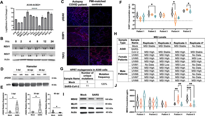
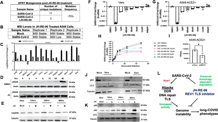
The repertoire of coronavirus disease 2019 (COVID-19)-mediated adverse health outcomes has continued to expand in infected patients, including the susceptibility to developing long-COVID; however, the molecular underpinnings at the cellular level are poorly defined. In this study, we report that SARS-CoV-2 (severe acute respiratory syndrome coronavirus 2) infection triggers host cell genome instability by modulating the expression of molecules of DNA repair and mutagenic translesion synthesis. Further, SARS-CoV-2 infection causes genetic alterations, such as increased mutagenesis, telomere dysregulation, and elevated microsatellite instability (MSI). The MSI phenotype was coupled to reduced MLH1, MSH6, and MSH2 in infected cells. Strikingly, pre-treatment of cells with the REV1-targeting translesion DNA synthesis inhibitor, JH-RE-06, suppresses SARS-CoV-2 proliferation and dramatically represses the SARS-CoV-2-dependent genome instability. Mechanistically, JH-RE-06 treatment induces autophagy, which we hypothesize limits SARS-CoV-2 proliferation and, therefore, the hijacking of host-cell genome instability pathways. These results have implications for understanding the pathobiological consequences of COVID-19.

Keywords: REV1; SARS-CoV-2; autophagy; microsatellite-instability; mutagenesis; telomeres.


Conflict of interest statement

Declarations Competing interests “P.Z. and J.H. are inventors of a patent on JH-RE-06 for cancer therapy.”

Figures

Figure 1
Figure 1. SARS-CoV-2 triggers genome instability.
(A) Relative expression of transcripts 48 hours post-SARS-CoV-2 infection (MOI 0.01) in A549-ACE2+ cells via a qPCR; n=9. (B) Western blots show change in REV1 and REV7 expression over time in A549-ACE2+ cells after SARS-CoV-2 infection (MOI 0.1); n=3. (C) Immunohistochemical images of 53 BP1, gH2AX, and TRF2 in lung tissues of autopsy COVID-19 patients and PMI (post-mortem interval) matched control lung tissues. (D-E) Western blots and their quantified plots of gH2AX expression in SARS-CoV-2 infected hamsters over time; n=3. (F) Relative concentration of 53BP1 in sera of long-COVID patients at three intervals, six months apart in an ELISA; n=3 technical replicates. (G) Table summarizes unique genetic alterations at exon 6 of the HPRT gene in A549-ACE2+ cells infected with SARS-CoV-2 for 48 hours at MOI 0.01; 2 biological and 5 technical replicates. (H) Table summarizes microsatellite instability in SARS-CoV-2-infected A549-ACE2+ cells (MOI 0.01) and in lung tissues of autopsy COVID-19 patients; n=3–4. (I) Western blots show MSH2, MLH1 and MSH6 expression in mock and SARS-CoV-2 infected (MOI 0.01) A549-ACE2+ cells; n=4. (J) Relative concentration of MSH2 in sera of long-COVID patients at three intervals, six months apart in an ELISA; n=5 technical replicates. Error bars represent standard error of the mean. ****P<0.0001, ***P<0.0001, **P<0.001, *P<0.01, statistical analysis noted in methods.
Figure 2
Figure 2. REV1 inhibitor JH-RE-06 suppresses SARS-CoV-2-dependent genome instability by triggering autophagy.
(A) Table summarizes unique genetic alterations at exon 6 of the HPRT gene in JH-RE-06-treated A549-ACE2+ cells infected with SARS-CoV-2 for 48 hours at MOI 0.01; 2 biological and 5 technical replicates; n=5–12. (B) Table summarizes microsatellite instability in JH-RE-06-treated A549-ACE2+ cells infected with SARS-CoV-2 for 48 hours at MOI 0.01; n=3. (C) Relative expression of transcripts 48 hours post-SARS-CoV-2 infection (MOI 0.01) in JH-RE-06-treated A549-ACE2+ cells via qPCR; n=3. (D and E) Western blots show gH2AX and CASP9 in a time course post-SARS-CoV-2 infection (MOI 0.01) in JH-RE-06-treated A549-ACE2+ cells; n=3. (F and G) Relative expression of SARS-CoV-2 N mRNA 48 hours post-infection (MOI 0.01) in JH-RE-06-treated Vero and A549-ACE2+ cells via a qPCR; n=3. (H) Relative expression of SARS-CoV-2 N mRNA over a time course of infection (MOI 0.01) in JH-RE-06-treated (5 and 10 mM) wildtype (WT) and STAT1 knockout (KO) A549-ACE2+ cells via a qPCR; n=3. (I) Plaque assay of supernatants from A549-ACE2+ cells treated with siRNAs against a non-targeting control or REV1 and infected with SARS-CoV-2; n=3. (J and K) Western blot shows expression of p65, p62, and LC3 in SARS-CoV-2 infected (MOI 0.1) A549-ACE2+ cells at 0, 12, 24 and 48 hours and treated with and without 1 mM of JH-RE-06; n=3. (L) Model shows SARS-CoV-2 dependent genome instability. Error bars represent standard error of the mean. ****P<0.0001, statistical analysis noted in methods.

References

    1. Taquet M.; Dercon Q.; Luciano S.; Geddes J. R.; Husain M.; Harrison P. J., Incidence, co-occurrence, and evolution of long-COVID features: A 6-month retrospective cohort study of 273,618 survivors of COVID-19. PLoS Med 2021, 18 (9), e1003773. - PMC - PubMed
    1. Douaud G.; Lee S.; Alfaro-Almagro F.; Arthofer C.; Wang C.; McCarthy P.; Lange F.; Andersson J. L. R.; Griffanti L.; Duff E.; Jbabdi S.; Taschler B.; Keating P.; Winkler A. M.; Collins R.; Matthews P. M.; Allen N.; Miller K. L.; Nichols T. E.; Smith S. M., SARS-CoV-2 is associated with changes in brain structure in UK Biobank. Nature 2022. - PMC - PubMed
    1. Xie Y.; Xu E.; Bowe B.; Al-Aly Z., Long-term cardiovascular outcomes of COVID-19. Nat Med 2022, 28 (3), 583–590. - PMC - PubMed
    1. Mavrikaki M.; Lee J. D.; Solomon I. H.; Slack F. J., Severe COVID-19 induces molecular signatures of aging in the human brain. medRxiv 2021. - PubMed
    1. Lee S.; Yu Y.; Trimpert J.; Benthani F.; Mairhofer M.; Richter-Pechanska P.; Wyler E.; Belenki D.; Kaltenbrunner S.; Pammer M.; Kausche L.; Firsching T. C.; Dietert K.; Schotsaert M.; Martinez-Romero C.; Singh G.; Kunz S.; Niemeyer D.; Ghanem R.; Salzer H. J. F.; Paar C.; Mulleder M.; Uccellini M.; Michaelis E. G.; Khan A.; Lau A.; Schonlein M.; Habringer A.; Tomasits J.; Adler J. M.; Kimeswenger S.; Gruber A. D.; Hoetzenecker W.; Steinkellner H.; Purfurst B.; Motz R.; Di Pierro F.; Lamprecht B.; Osterrieder N.; Landthaler M.; Drosten C.; Garcia-Sastre A.; Langer R.; Ralser M.; Eils R.; Reimann M.; Fan D. N. Y.; Schmitt C. A., Virus-induced senescence is a driver and therapeutic target in COVID-19. Nature 2021, 599 (7884), 283–289. - PubMed

Publication types

LinkOut - more resources