[Preprint]. 2022 Apr 11:rs.3.rs-1493296.
doi: 10.21203/rs.3.rs-1493296/v1.

Famotidine activates the vagus nerve inflammatory reflex to attenuate cytokine storm

Affiliations

Famotidine activates the vagus nerve inflammatory reflex to attenuate cytokine storm

Huan Yang et al. Res Sq. .

Update in

Abstract

Background. Severe COVID-19 is characterized by pro-inflammatory cytokine release syndrome (cytokine storm) which causes high morbidity and mortality. Recent observational and clinical studies suggest famotidine, a histamine 2 receptor (H2R) antagonist widely used to treat gastroesophageal reflux disease , attenuates the clinical course of COVID-19. Because evidence is lacking for a direct antiviral activity of famotidine, a proposed mechanism of action is blocking the effects of histamine released by mast cells. Here we hypothesized that famotidine activates the inflammatory reflex, a brain-integrated vagus nerve mechanism which inhibits inflammation via alpha 7 nicotinic acetylcholine receptor ( α7nAChR ) signal transduction, to prevent cytokine storm. Methods. The potential anti-inflammatory effects of famotidine and other H2R antagonists was assessed in mice exposed to lipopolysaccharide (LPS)-induced cytokine storm. As the inflammatory reflex is integrated and can be stimulated in the brain, and H2R antagonists penetrate the blood brain barrier poorly, famotidine was administered by intracerebroventricular (ICV) or intraperitoneal (IP) routes. Results. Famotidine administered IP significantly reduced serum and splenic LPS-stimulated tumor necrosis factor α and interleukin-6 concentrations, significantly improving survival. The effects of ICV famotidine were significantly more potent as compared to the peripheral route. Mice lacking mast cells by genetic deletion also responded to famotidine, indicating the anti-inflammatory effects are not mast cell dependent. Either bilateral sub-diaphragmatic vagotomy or genetic knock-out of α7nAChR abolished the anti-inflammatory effects of famotidine, indicating the inflammatory reflex as famotidine's mechanism of action. While the structurally similar H2R antagonist tiotidine displayed equivalent anti-inflammatory activity, the H2R antagonists cimetidine or ranitidine were ineffective even at very high dosages. Conclusions. These observations reveal a previously unidentified vagus nerve-dependent anti-inflammatory effect of famotidine in the setting of cytokine storm which is not replicated by high dosages of other H2R antagonists in clinical use. Because famotidine is more potent when administered intrathecally, these findings are also consistent with a primarily central nervous system mechanism of action.


Conflict of interest statement

Competing interests

HY, SSC and KJT are co-inventors of a patent application (Role of the central nervous nerve system and vagus nerve signaling in famotidine-mediated anti-inflammatory effects). All other authors declare no competing interests.

Figures

Figure 1
Figure 1
Famotidine attenuates lipopolysaccharide (LPS)-induced in ammatory responses in mice. A-D. Male C57BL/6 mice, 8–12 weeks of age, were injected with LPS (7 mg/kg) with or without famotidine (FM, 0.4 or 4 mg/kg, in 100 μl volume), intraperitoneally (IP) 30 min before LPS injection. Mice were euthanized 2.5 hours after LPS administration and serum and spleen TNF and IL-6 were measured. N=10 mice per group. *P<0.01, **P=0.001, ***P<0.001. E. Male C57BL/6 mice, 8–12 weeks old, were injected with LPS (6mg/kg, IP). Famotidine (FM, 4 mg/kg) or PBS (in 100 μl volume) were injected intraperitoneally twice a day for 3 days, survival was monitored for 2 weeks. N= 30 mice per group. *P=0.001 vs. LPS+PBS group.
Figure 2
Figure 2
Famotidine is more potent when administered into the central nervous system. A-D. Famotidine (intracerebroventrical, ICV) injection attenuated LPS-induced systemic TNF and IL-6 release in mice. Male C57BL/6 mice had ICV injection of PBS or famotidine (0.04 or 0.4 mg/kg in 5 μl volume) 30 min before LPS. LPS was administered IP at 7 mg/kg. Mice were euthanized 2.5 hours post-LPS injection and serum and spleen were harvested for analyses. N=5 for normal group. N=10 for FM alone. N=13 or 14 for other groups. *P=0.0004. **P=0.0001. ***P<0.0001.
Figure 3
Figure 3
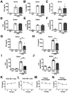
Mast cell de cient KitW-sh/KitW-sh sash mice had similar LPS-induced TNF, IL-6, IL-1β response as wild type mice. Male wild type (WT) or “sash” mice received LPS injection (IP, 7mg/kg) and were euthanized 2.5 hours post-LPS injection. Serum and spleen TNF, IL-6 and IL-1β were measured. N=3. G-J Famotidine, administered ICV, reduced LPS-induced TNF and IL-6 release in mast cell de cient KitW-sh/KitW-sh sash mice at 2.5 hours post LPS exposure. Male KitW-sh/KitW-sh sash mice received famotidine or PBS (0.4 mg/kg, in 5 μl volume) administered ICV at 30 min prior to LPS injection (IP, 7mg/kg). Mice were euthanized 2.5 hours post-LPS injection and serum TNF and IL-6 were measured. N=3 for normal wild type, n= 4 for normal “sash”, 5 for LPS, 5 or 7 for LPS+ famotidine group. *P<0.001. **P<0.0001. K-N. RAW 264.7 cells (K-L) or thioglycollate-elicited mouse primary peritoneal macrophages (M-N) in 96-well culture plates were stimulated with LPS (0.4ng/ml) in combination with various amounts of famotidine for 16 hours. TNF (K,M) and IL-6 (L,N) released in the supernatants were measured. n = 3–6 per treatment. *P<0.05 vs. LPS alone.
Figure 4
Figure 4
Famotidine activates the vagus nerve. A-B: Representative recordings of the vagus nerve signals in wild type mice pre- (baseline) and post- (A) saline or (B) famotidine administration ICV. Data is representative of 4 or 5 animals per group. Total spike count during recordings over the entire 4-minute post-ICV administration of saline and famotidine. (n=4 for saline and 5 for famotidine). C: *P = 0.029.
Figure 5
Figure 5
The suppressive effects of famotidine on LPS-induced pro-in ammatory cytokine release were abrogated in bilateral sub-diaphragm vagotomized mice. A-D. Male C57BL/6 mice had vagotomy (VGX; bilateral sub-diaphragmatic, with transgastric pyloric dilation to reduce stomach distension) and recovered for 7 days. Mice received famotidine (4 mg/kg) IP 30 min prior to LPS (IP, 7mg/kg). Mice were euthanized 2.5 hours later, serum and spleen TNF and IL-6 were measured. N=5 for normal; 7 per group for others.
Figure 6
Figure 6
Suppressive effects of famotidine on LPS-induced TNF and IL-6 release were greatly attenuated and mortality increased in α7 nicotinic acetylcholine receptor knockout mice. A-D. Male C57BL/6 or α7nAChR knockout mice (α7KO; 8–12 weeks of age) received famotidine or PBS (0.4 mg/kg, in 5 μl volume) administered ICV at 30 min prior to LPS injection (IP, 7mg/kg). Mice were euthanized 2.5 hours post-LPS injection, serum and spleen TNF (B-C) and IL-6 (D-E) were measured. N=7 for wild type and 6 for α7nAChR KO controls, 9–11 per group for others. E. Male α7nAChR knockout mice, 8–12 weeks old, were injected with LPS (6mg/kg, IP). Famotidine (FM) or PBS (4 mg/kg, in 100 μl volume) were injected intraperitoneally twice a day for 3 days, survival was monitored for 2 weeks. N=17 for PBS control and 18 for FM mice per group.

References

    1. Mehta P, et al. (2020) COVID-19: consider cytokine storm syndromes and immunosuppression. Lancet (London, England) 395: 1033–1034. - PMC - PubMed
    1. Wang J, Jiang M, Chen X, Montaner LJ. (2020) Cytokine storm and leukocyte changes in mild versus severe SARS-CoV-2 infection: Review of 3939 COVID-19 patients in China and emerging pathogenesis and therapy concepts. Journal of leukocyte biology 108: 17–41. - PMC - PubMed
    1. Jafarzadeh A, Chauhan P, Saha B, Jafarzadeh S, Nemati M. (2020) Contribution of monocytes and macrophages to the local tissue inflammation and cytokine storm in COVID-19: Lessons from SARS and MERS, and potential therapeutic interventions. Life sciences 257: 118102. - PMC - PubMed
    1. Moore JB, June CH. (2020) Cytokine release syndrome in severe COVID-19. Science 368: 473–474. - PubMed
    1. Sharun K, Tiwari R, Dhama J, Dhama K. (2020) Dexamethasone to combat cytokine storm in COVID-19: Clinical trials and preliminary evidence. Int J Surg 82: 179–181. - PMC - PubMed

Publication types