. 2022 Jul 29;145(7):2407-2421.
doi: 10.1093/brain/awac121.

Cortical phase-amplitude coupling is key to the occurrence and treatment of freezing of gait

Affiliations

Cortical phase-amplitude coupling is key to the occurrence and treatment of freezing of gait

Zixiao Yin et al. Brain. .

Abstract

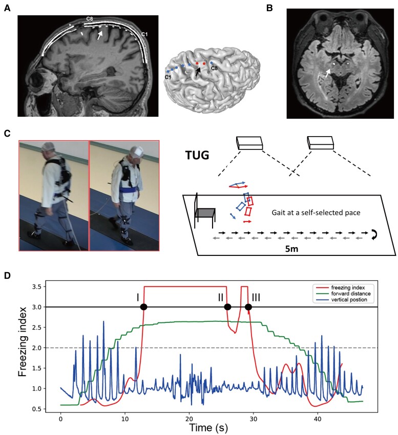
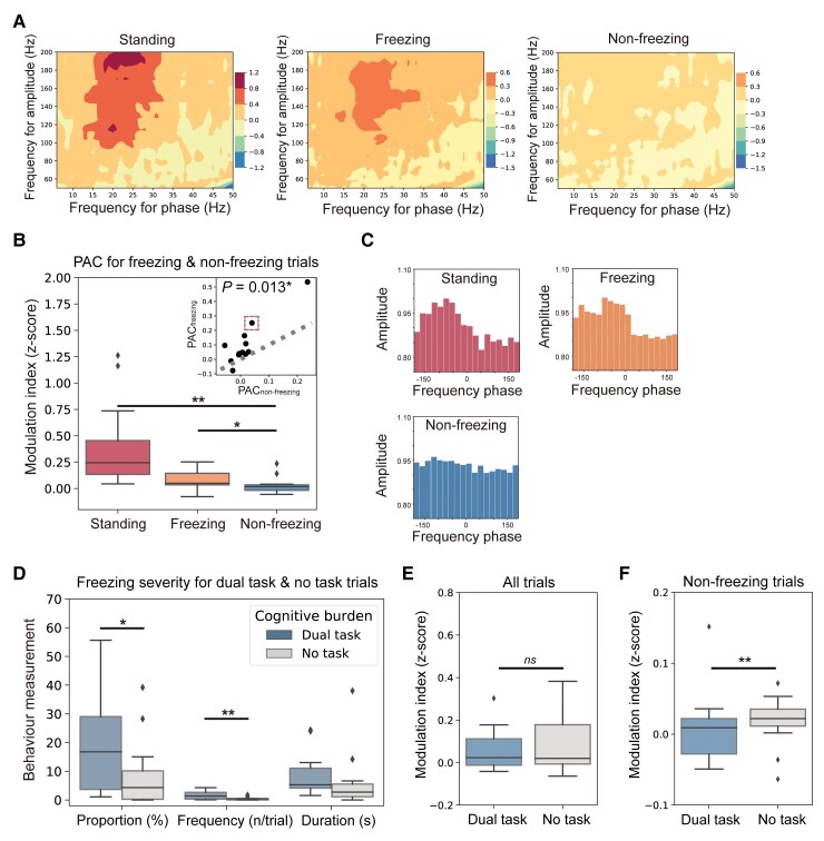
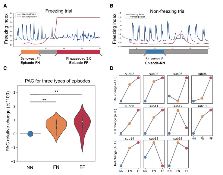
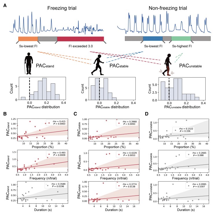
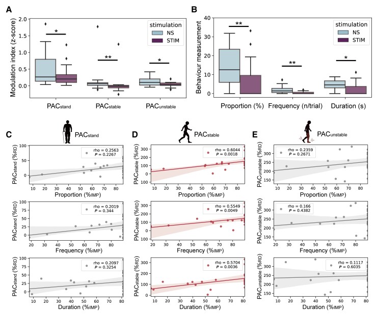
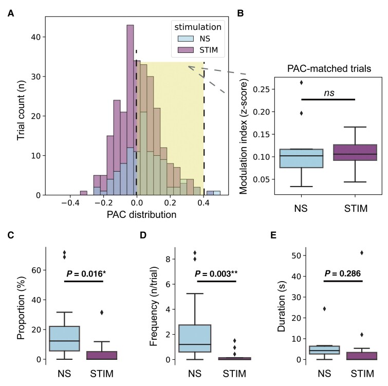
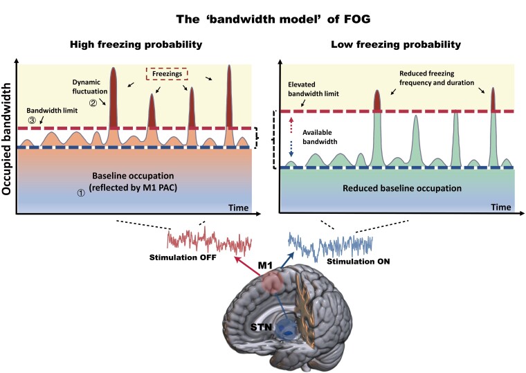
Freezing of gait is a debilitating symptom in advanced Parkinson's disease and responds heterogeneously to treatments such as deep brain stimulation. Recent studies indicated that cortical dysfunction is involved in the development of freezing, while evidence depicting the specific role of the primary motor cortex in the multi-circuit pathology of freezing is lacking. Since abnormal beta-gamma phase-amplitude coupling recorded from the primary motor cortex in patients with Parkinson's disease indicates parkinsonian state and responses to therapeutic deep brain stimulation, we hypothesized this metric might reveal unique information on understanding and improving therapy for freezing of gait. Here, we directly recorded potentials in the primary motor cortex using subdural electrocorticography and synchronously captured gait freezing using optoelectronic motion-tracking systems in 16 freely-walking patients with Parkinson's disease who received subthalamic nucleus deep brain stimulation surgery. Overall, we recorded 451 timed up-and-go walking trials and quantified 7073 s of stable walking and 3384 s of gait freezing in conditions of on/off-stimulation and with/without dual-tasking. We found that (i) high beta-gamma phase-amplitude coupling in the primary motor cortex was detected in freezing trials (i.e. walking trials that contained freezing), but not non-freezing trials, and the high coupling in freezing trials was not caused by dual-tasking or the lack of movement; (ii) non-freezing episodes within freezing trials also demonstrated abnormally high couplings, which predicted freezing severity; (iii) deep brain stimulation of subthalamic nucleus reduced these abnormal couplings and simultaneously improved freezing; and (iv) in trials that were at similar coupling levels, stimulation trials still demonstrated lower freezing severity than no-stimulation trials. These findings suggest that elevated phase-amplitude coupling in the primary motor cortex indicates higher probabilities of freezing. Therapeutic deep brain stimulation alleviates freezing by both decoupling cortical oscillations and enhancing cortical resistance to abnormal coupling. We formalized these findings to a novel 'bandwidth model,' which specifies the role of cortical dysfunction, cognitive burden and therapeutic stimulation on the emergence of freezing. By targeting key elements in the model, we may develop next-generation deep brain stimulation approaches for freezing of gait.

Keywords: Parkinson’s disease; deep brain stimulation; freezing of gait; motor cortex; phase amplitude coupling.


Figures

Figure 1
Figure 1
Electrode localization, experimental setup and representation of the freezing index. (A) Localization of electrocorticography (ECoG) electrodes. The eight contacts (C1–C8) are visualized in the merged image of preoperative MRI and postoperative CT (left). C8 is the contact closest to the DBS bone hole. The white arrow points to the primary motor cortex. A reconstruction of the cortex and the eight contacts relative to the primary motor cortex (black arrow) is shown in the right figure. (B) Localization of the STN electrodes (white arrow) in the merged image of preoperative MRI and postoperative CT. (C) Experimental setup and protocol. Patients were asked to walk barefoot while completing a 10 m (5 m one way) back-and-forth timed up-and-go task at a self-selected pace with sensors attached to the lower limbs. The instant coordinates of the sensor were captured through an optoelectronic motion tracking system hanging on walls on both sides. Synchronized ECoG potentials were recorded through an extended cable. (D) The representative diagram of the freezing index (FI). The blue line represents the vertical position of the foot. The green line represents the forward position of the foot. The red line represents the FI. When the vertical kinematic rhythm becomes irregular and the forward motion stagnates, FI rises and exceeds the 3-point threshold (solid black line). Notably, if the FI drops below ‘3’ but then rises back, with the lowest value still over ‘2’ (grey dashed line), we consider this as one continuous freezing event rather than two. Thus, the diagram shows one continuous freezing event lasting from time point I to time point III. Because FI does not drop below ‘2’, time point II does not mark the end of this freezing event.
Figure 2
Figure 2
Freezing trials have higher M1 PAC than non-freezing trials. (A) Co-modulograms showing group-level M1 beta-gamma PAC in rest standing (left), freezing (middle) and non-freezing (right) trials. Deep colours indicate high PAC. (B) Box plots indicating the comparison of PAC between rest standing, freezing and non-freezing trials, which was tested using the Wilcoxon signed-rank test. The top right plot shows the paired-comparison results. Each dot represents a patient. Dots landed above the grey dashed line have higher PACs in freezing trials (PACfreezing). Dots landed below the grey dashed line have higher PACs in non-freezing trials (PACnon-freezing). (C) Examples show the distributions of amplitude and preferred phase of the coupling in rest standing (red), freezing (orange) and non-freezing trials (blue). These data are based on Patient Sub8, which is represented by the dot marked with a red dashed box in B (top right plot). (D) Box plots comparing freezing time proportion, freezing frequency and duration per freezing between dual-tasking and no-task trials. (E) Box plots comparing PAC between dual-tasking and no-task conditions in all trials. (F) Box plots comparing PAC between dual-tasking and no-task conditions in non-freezing trials. In box plots, the lower and upper borders of the box represent the 25th and 75th percentiles, respectively. The centreline represents the median. The whiskers extend to the smallest and largest data-points that are not outliers (1.5 times the interquartile range). Significant P-values after Bonferroni correction are indicated. **P < 0.01, *P < 0.05, signed-rank test.
Figure 3
Figure 3
Non-freezing episodes in freezing trials also have higher PAC in M1. (A) Schematic diagram depicting the slicing of non-freezing episodes (marked in orange, FN) and freezing episodes (marked in red, FF) in freezing trials. The blue line represents the vertical position of the foot, and the red line represents the freezing index (FI). (B) Schematic diagram depicting the slicing of normal-walking episodes (marked in blue, NN) in non-freezing trials. (C) Violin plots indicate the comparison of relative PAC change between FN, FF and NN episodes. The relative change was calculated as the percentage change with respect to NN scaling to the max value. Violin plots outline illustrate kernel probability density, with overlaid box plots using the same conventions as in Fig. 2B. (D) A similar neurophysiological pattern that was characterized by higher M1 PAC in FN and FF episodes was presented in all subjects. **P < 0.01, signed-rank test.
Figure 4
Figure 4
PAC during stable walking is correlated with freezing severity. (A) Distribution of condition-wise PACs during pre-walking standing (PACstand, left), stable walking (PACstable, middle) and unstable walking (PACunstable, right). (B) Regression plots showing the correlation between PACstand and the freezing time proportion (top), freezing frequency (middle) and duration per freezing (bottom). (C) Regression plots showing the correlation between PACstable and the freezing time proportion (top), freezing frequency (middle) and duration per freezing (bottom). (D) Regression plots showing the correlation between PACunstable and the freezing time proportion (top), freezing frequency (middle) and duration per freezing (bottom). Note that each patient has three data points resulting in 14 × 3 PAC values (n = 42), as PAC was calculated in three stimulation conditions (i.e. HFS, LFS and no-stimulation). Statistical dependence within subjects was accounted for using linear mixed-effects models. Significant correlations after Bonferroni correction are marked in red.
Figure 5
Figure 5
The reduction of PACstable predicts the improvement of freezing severity induced by DBS. (A) Box plots comparing PACstand, PACstable and PACunstable between no-stimulation (NS) and stimulation (STIM) conditions. (B) Box plots comparing freezing time proportion, freezing frequency and duration per freezing between NS and STIM conditions. Same conventions as in Fig. 2B. **P < 0.01, *P < 0.05, signed-rank test. (C) Regression plots showing the correlation between the percentage change of PACstand and the percentage change of freezing time proportion (top), freezing frequency (middle) and duration per freezing (bottom). (D) Regression plots showing the correlation between the percentage change of PACstable and the percentage change of freezing time proportion (top), freezing frequency (middle) and freezing duration (bottom). (E) Regression plots showing the correlation between the percentage change of PACunstable and the percentage change of freezing time proportion (top), freezing frequency (middle) and duration per freezing (bottom). Note that each patient has two data points resulting in 14 × 2 PAC values (n = 28), as the reduction of PAC was calculated in two stimulation conditions (i.e. HFS and LFS). Statistical dependence within subjects was accounted for using linear mixed effects models. Significant correlations after Bonferroni correction are marked in red. %RD = percentage reduction; %IMP = percentage improvement.
Figure 6
Figure 6
Stimulation trials have lower freezing severity than no-stimulation trials even when under similar levels of PAC. (A) The distribution of PAC in no-stimulation (NS) and stimulation (STIM) trials. Trials with PACs between 0 and 0.4 in both the NS and STIM groups were picked out as PAC-matched trials (marked by yellow shadow background) for further analyses. Box plots showing the (B) comparison of PAC, (C) comparison of freezing time proportion, (D) comparison of freezing frequency and (E) comparison of duration per freezing between PAC-matched NS and STIM trials. Same conventions as in Fig. 2B. **P < 0.01, *P < 0.05, signed-rank test. ns = not significant.
Figure 7
Figure 7
A graphical representation of the proposed ‘bandwidth model’ of FOG. Three main elements constitute the model: (i) the baseline occupation; (ii) the dynamic fluctuation; and (iii) the bandwidth limit. The x-axis represents the time axis, and y-axis represents the occupied bandwidth. When baseline occupation plus dynamic fluctuation exceeds the bandwidth limit, freezing occurs. Baseline occupation can be quantified through M1 PAC. In the off-stimulation state (left), the baseline occupation (M1 PAC) maintains at a high level, leading to a high probability of exceeding the bandwidth limit. In the on-stimulation state (right), a reduction of baseline occupation and an elevation of bandwidth limit clean up larger available bandwidth that can be used to process dynamic fluctuation, leading to a lower probability of freezing.

Comment in

References

    1. Nutt JG, Bloem BR, Giladi N, Hallett M, Horak FB, Nieuwboer A. Freezing of gait: moving forward on a mysterious clinical phenomenon. Lancet Neurol. 2011;10(8):734–744. - PMC - PubMed
    1. Moore O, Peretz C, Giladi N. Freezing of gait affects quality of life of peoples with Parkinson’s disease beyond its relationships with mobility and gait. Mov Disord. 2007;22(15):2192–2195. - PubMed
    1. Muslimovic D, Post B, Speelman JD, Schmand B, de Haan RJ; CARPA Study Group . Determinants of disability and quality of life in mild to moderate Parkinson disease. Neurology. 2008;70(23):2241–2247. - PubMed
    1. Fasano A, Daniele A, Albanese A. Treatment of motor and non-motor features of Parkinson’s disease with deep brain stimulation. Lancet Neurol. 2012;11(5):429–442. - PubMed
    1. Bohnen NI, Costa RM, Dauer WT, et al. . Discussion of research priorities for gait disorders in Parkinson’s disease. Mov Disord. 2022;37(2):253–263. - PMC - PubMed

Publication types

MeSH terms