

Effectiveness of Ilizarov Ankle Arthrodesis in the Treatment of End-Stage Varus Ankle Osteoarthritis: A Retrospective Study
- PMID: 35441475
- PMCID: PMC9087455
- DOI: 10.1111/os.13286
Effectiveness of Ilizarov Ankle Arthrodesis in the Treatment of End-Stage Varus Ankle Osteoarthritis: A Retrospective Study
Abstract
Objective: To evaluate the outcomes of Ilizarov ankle arthrodesis in the treatment of end-stage varus ankle osteoarthritis (OA).
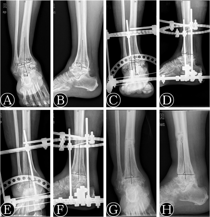
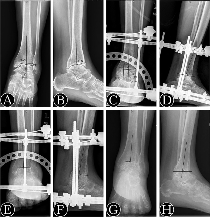
Methods: This was a retrospective study of 63 patients with varus ankle OA who underwent Ilizarov ankle arthrodesis between June 2013 and December 2018. There were 24 males and 39 females with an average age of 56.57 ± 4.45 years (range, 47-64 years). Thirty-six cases were affected on the left side, and 27 were affected on the right side. The patients' mean body mass index (BMI) was 25.18 ± 2.93 kg/m2 . According to the modified Takakura staging criteria, there were 18 cases of stage 3b (28.57%) and 45 cases of stage 4 (71.43%). Nine patients were primary (14.29%), 48 were traumatic (76.19%), and six were caused by rheumatoid OA (9.52%). Functional assessments were performed according to the American Orthopedic Foot and Ankle Society (AOFAS) ankle-hindfoot score, Ankle Osteoarthritis Scale (AOS), and visual analogue scale (VAS). The tibial anterior surface angle (TAS), coronal plane tibial-talar angle (CPT), talar tilt angle (TT), deformity angle (DA), and tibial lateral surface angle (TLS) were assessed on X-ray films.
Results: The average operation time was 147.84 ± 13.67 min (range, 135-168 min). The average follow-up time was 34.24 ± 8.72 months (range, 24-61 months). Bony fusion was achieved in all ankles, and the fusion time was 12.43 ± 1.99 weeks on average. The average AOFAS score at the final follow-up increased from 42.14 ± 8.66 to 80.90 ± 6.80. The average VAS score and AOS pain and disability scores at the final follow-up decreased from 7.29 ± 1.27 to 2.24 ± 0.94, from 67.94 ± 7.68 to 27.92 ± 5.82, and from 71.64 ± 9.37 to 41.32 ± 8.99, respectively. The average TAS, CPT, and TLS at the final follow-up increased from 77.76° ± 4.44° to 89.81° ± 1.25°, from 69.04° ± 3.73° to 90.43° ± 1.80°, and from 82.14° ± 3.77° to 88.67° ± 2.50°, respectively. The average TT and DA at the final follow-up decreased from 8.76° ± 4.30° to 2.05° ± 1.28° and from 20.95° ± 3.73° to 1.57° ± 0.93°, respectively. Three patients developed superficial pin tract infections, all settled with local dressing and antibiotic treatment. Two patients were found to have subtalar arthritis and underwent conservative treatment.
Conclusion: Ankle arthrodesis using the Ilizarov technique is efficient in treating end-stage varus ankle OA.
Keywords: Ankle arthrodesis; Ankle osteoarthritis; Deformity correction; Ilizarov; Varus deformity.
© 2022 The Authors. Orthopaedic Surgery published by Tianjin Hospital and John Wiley & Sons Australia, Ltd.
Figures


References
-
- Hongmou Z, Xiaojun L, Yi L, Hongliang L, Junhu W, Cheng L. Supramalleolar osteotomy with or without fibular osteotomy for varus ankle arthritis. Foot Ankle Int. 2016;37:1001–7. - PubMed
-
- Hennessy MS, Molloy AP, Wood EV. Management of the varus arthritic ankle. Foot Ankle Clin. 2008;13:417‐442. - PubMed
MeSH terms
Grants and funding
- 2022SCUH0014/"Zero to One" Innovation Research Project of Sichuan University
- 2021M692279/China Postdoctoral Science Foundation
- Z20192013/National Clinical Research Center for Geriatrics, West China Hospital, Sichuan University
- 81874002/National Natural Science Foundation of China
- 2019HXBH068/West China hospital postdoctoral research and development fund
LinkOut - more resources
Full Text Sources
Medical
