. 2022 Mar 21;92(S4):e2021352.
doi: 10.23750/abm.v92iS4.12823.

Far Lateral Approach

Affiliations

Far Lateral Approach

Sabino Luzzi et al. Acta Biomed. .

Abstract

The far lateral approach is an inferolateral extension of the lateral suboccipital approach. Designed for clipping of the aneurysms of the vertebrobasilar junction and proximal segments of the posterior inferior cerebellar artery, it became over the years a workhorse approach for ventral foramen magnum meningiomas and other intradural lesions located anterior to the dentate ligament. This article summarizes the technical key aspects of the far lateral approach and transcondylar, supracondylar, and paracondylar extension.


Figures
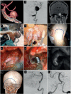
Figure 1.
Figure 1.. Layer-by-layer dissection of the nuchal muscles. (A) Skin incision. First (B), second (C), third (D), fourth (E) muscular layer. IO: inferior oblique muscle; LC: longissimus capitis muscle; OA: occipital artery; PT: posterior triangle of the neck; RC Maj: rectus capitis posterior major muscle; SCM: sternocleidomastoid muscle; Sem. C: semispinalis capitis muscle; Sem. C: semispinalis capitis muscle; SO: superior oblique muscle; Spl. C: splenius capitis muscle; T: trapezius muscle.
Figure 2.
Figure 2.. (A, B) Far lateral approach. Transcondylar (C, D), supracondylar (E, F), and paracondylar (G, H) extension of the far lateral approach as regards craniotomy (left column) and intradural exposure (right column). IHT: infrahypoglossal triangle; SHT: suprahypoglossal triangle; VAT: vagoaccessory triangle.
Figure 3.
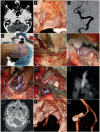
Figure 3.. Axial contrast-enhanced CT scan (A) and CT angiography (B). (C) Digital subtraction angiography of the left vertebral artery in anterior-posterior projection. Right modified park-bench position (D), dura opening (E), and aneurysm exposure (F). (G, H) Clipping of the aneurysm. (I) Indocyanine green videoangiography after clipping. (J) Postoperative axial bone window CT scan. (K, L) Postoperative 3D volume rendering CT angiography.
Figure 4.
Figure 4.. (A) Preoperative 3D volume rendering CT angiography; (B) Digital subtraction angiography of the left vertebral artery in the lateral projection; (C) Coronal contrast-enhanced MRI. (D) Patient in right modified park-bench position. (E) Neurophysiologi-cal monitoring. (F) Reverse hockey stick incision. (G, H) Clipping of the aneurysm. (I) Indocyanine green videoangiography after clipping. (J) Postoperative 3D CT scan. (K) Postoperative digital subtraction angiography of the left vertebral artery in lateral (K) and anterior-posterior (L) projection with the double catheter approach.

References

    1. Heros RC. Lateral suboccipital approach for vertebral and vertebrobasilar artery lesions. J Neurosurg. 1986;64(4):559–562. - PubMed
    1. Bruneau M, George B. Classification system of foramen magnum meningiomas. J Craniovertebr Junction Spine. 2010;1(1):10–7. - PMC - PubMed
    1. Day JD, Fukushima T, Giannotta SL. Cranial base approaches to posterior circulation aneurysms. J Neurosurg. 1997;87(4):544–554. - PubMed
    1. Bertalanffy H, Seeger W. The dorsolateral, suboccipital, transcondylar approach to the lower clivus and anterior portion of the craniocervical junction. Neurosurgery. 1991;29(6):815–821. - PubMed
    1. de Oliveira E, Rhoton AL,, Jr, Peace D. Microsurgical anatomy of the region of the foramen magnum. Surg Neurol. 1985;24(3):293–352. - PubMed

LinkOut - more resources