

Review
doi: 10.23750/abm.v92iS4.12784.
Cranio-Orbito-Zygomatic Approach
Affiliations
- PMID: 35441603
- PMCID: PMC9179061
- DOI: 10.23750/abm.v92iS4.12784
Item in Clipboard
Review
Cranio-Orbito-Zygomatic Approach
Acta Biomed.
.
Abstract
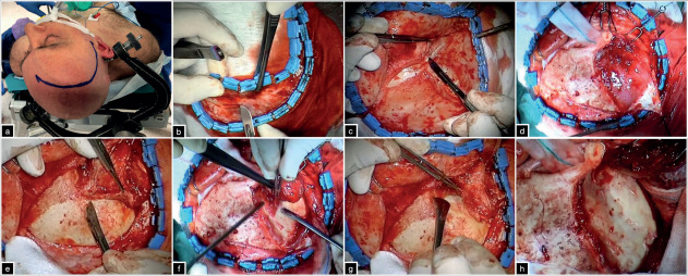
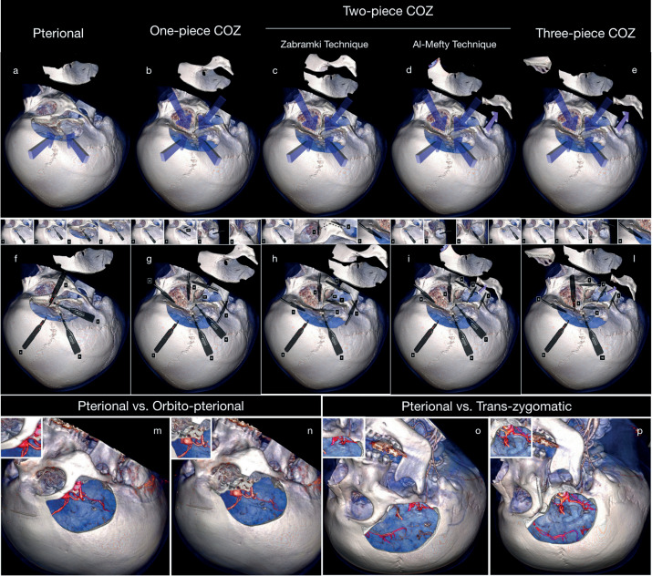
The cranio-orbito-zygomatic (COZ) approach consists of an extension of the pterional approach characterized by the removal of the superolateral part of the orbital rim and zygoma. This key step tremendously increases the angular exposure to some deep targets and overall surgical freedom to the lesion. In this article we review the technical variations of the COZ approach, mainly focusing on the differential quantitative effects coming from the orbital osteotomy compared to the zygomatic one.
Figures
References
-
- Pellerin P, Lesoin F, Dhellemmes P, Donazzan M, Jomin M. Usefulness of the orbitofrontomalar approach associated with bone reconstruction for frontotemporosphenoid meningiomas. Neurosurgery. 1984;15(5):715–718. - PubMed
-
- Hakuba A, Liu S, Nishimura S. The orbitozygomatic infratemporal approach: a new surgical technique. Surg Neurol. 1986;26(3):271–276. - PubMed
-
- Al-Mefty O. Supraorbital-pterional approach to skull base lesions. Neurosurgery. 1987;21(4):474–477. - PubMed
-
- Aziz KM, Froelich SC, Cohen PL, Sanan A, Keller JT, van Loveren HR. The one-piece orbitozygomatic approach: the MacCarty burr hole and the inferior orbital fissure as keys to technique and application. Acta Neurochir (Wien) 2002;144(1):15–24. - PubMed
-
- Delashaw JB,, Jr, Tedeschi H, Rhoton AL. Modified supraorbital craniotomy: technical note. Neurosurgery. 1992;30(6):954–956. - PubMed
Publication types
MeSH terms
LinkOut - more resources
Full Text Sources
