

Pterional Approach
- PMID: 35441604
- PMCID: PMC9179065
- DOI: 10.23750/abm.v92iS4.12775
Pterional Approach
Abstract
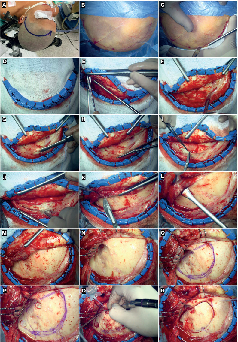
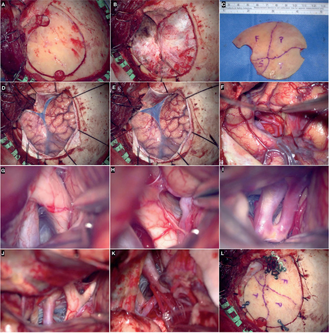
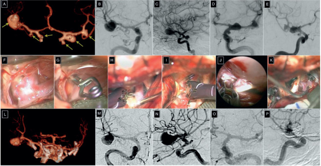
The pterional approach is a workhorse in neurosurgery, to the point where perfect knowledge of its execution is essential in neurosurgical daily practice. The pterional transsylvian corridor is used to treat aneurysms involving anterior circulation, basilar apex, the proximal segment of the superior cerebellar and posterior cerebral artery, arteriovenous malformations and cavernous hemangiomas of the basal forebrain, anterior and middle skull base tumors, gliomas of the frontal, parietal, and temporal opercula, insula, mediobasal temporal region, cerebral peduncles, interpeduncular fossa, and also orbital lesions. We herein overview the core technique and variations of the pterional approach aimed at broadening surgical freedom and decreasing the risk of approach-related complications.
Conflict of interest statement
Each author declares that he or she has no commercial associations (e.g. consultancies, stock ownership, equity interest, patent/licensing arrangement etc.) that might pose a conflict of interest in connection with the submitted article
Figures

References
-
- Yaargil M, Yaşargil M. Interfascial pterional (frontotem-porosphenoidal) craniotomy: Georg Thieme Verlag. 1984:217–20 p.
-
- Yaşargil M.G. FJL, Ray M.W. 'The Operative Approach to Aneurysms of the Anterior Communicating Artery. In: al. KHe, editor. Advances and Technical Standards in Neurosurgery, vol 2. Vienna: Springer. 1975
-
- Yasargil MG, Reichman MV, Kubik S. Preservation of the frontotemporal branch of the facial nerve using the inter-fascial temporalis flap for pterional craniotomy. Technical article. J Neurosurg. 1987;67(3):463–466. - PubMed
-
- Chaddad-Neto F, Doria-Netto HL, Campos-Filho JM, Ribas ES, Ribas GC, Oliveira E. Head positioning for anterior circulation aneurysms microsurgery. Arq Neuropsiqui-atr. 2014;72(11):832–840. - PubMed
-
- Oikawa S, Mizuno M, Muraoka S, Kobayashi S. Retrograde dissection of the temporalis muscle preventing muscle atrophy for pterional craniotomy. Technical note. J Neurosurg. 1996;84(2):297–299. - PubMed
Publication types
MeSH terms
LinkOut - more resources
Full Text Sources
Medical
Miscellaneous
