. 2022 Jul 4;34(7):2765-2784.
doi: 10.1093/plcell/koac114.

Fungi hijack a ubiquitous plant apoplastic endoglucanase to release a ROS scavenging β-glucan decasaccharide to subvert immune responses

Affiliations

Fungi hijack a ubiquitous plant apoplastic endoglucanase to release a ROS scavenging β-glucan decasaccharide to subvert immune responses

Balakumaran Chandrasekar et al. Plant Cell. .

Abstract

Plant pathogenic and beneficial fungi have evolved several strategies to evade immunity and cope with host-derived hydrolytic enzymes and oxidative stress in the apoplast, the extracellular space of plant tissues. Fungal hyphae are surrounded by an inner insoluble cell wall layer and an outer soluble extracellular polysaccharide (EPS) matrix. Here, we show by proteomics and glycomics that these two layers have distinct protein and carbohydrate signatures, and hence likely have different biological functions. The barley (Hordeum vulgare) β-1,3-endoglucanase HvBGLUII, which belongs to the widely distributed apoplastic glycoside hydrolase 17 family (GH17), releases a conserved β-1,3;1,6-glucan decasaccharide (β-GD) from the EPS matrices of fungi with different lifestyles and taxonomic positions. This low molecular weight β-GD does not activate plant immunity, is resilient to further enzymatic hydrolysis by β-1,3-endoglucanases due to the presence of three β-1,6-linked glucose branches and can scavenge reactive oxygen species. Exogenous application of β-GD leads to enhanced fungal colonization in barley, confirming its role in the fungal counter-defensive strategy to subvert host immunity. Our data highlight the hitherto undescribed capacity of this often-overlooked EPS matrix from plant-associated fungi to act as an outer protective barrier important for fungal accommodation within the hostile environment at the apoplastic plant-microbe interface.


Figures

Figure 1
Figure 1
Fungal EPS matrix revealed by the fluorescently labeled β-glucan binding lectin SiWSC3-His-FITC488 during root colonization. The β-glucan-binding SiWSC3-His and the chitin-binding WGA lectins were used as molecular probes to visualize the fungal EPS matrix and CW of S. indica and B. sorokiniana, respectively. Magenta pseudocolor corresponds to FITC488-labeled SiWSC3-His. Cyan pseudocolor corresponds to WGA-AF594. (A), (C), (E), and (G) are merged confocal microscopy images of SiWSC3-His-FITC488 and WGA-AF594. (B), (D), (F), and (H) display the EPS matrix of S. indica or B. sorokiniana stained by SiWSC3-His-FITC488 during colonization of Arabidopsis or barley roots. (A) and (B) show S. indica intracellular colonization of a barley root cell with abundant production of the β-glucan EPS matrix. The microscopy was repeated at least 10 times with two independent SiWSC3-His-FITC488 batches and independent Arabidopsis or barley plants colonized by S. indica or B. sorokiniana. The fungal matrix was not a sporadic observation but regularly observed with both fungi. WGA, wheat germ agglutinin.
Figure 2
Figure 2
Proteomics and glycosyl linkage analysis of S. indica EPS matrix and CW. A, Venn diagram of proteins identified in the EPS matrix, CW, and/or culture filtrate from three cultivation media (CM, YPD, and TSB). B, Proteins with WSC domain/s show higher abundances in the EPS matrix of S. indica (see also Supplemental Figure S2 and Supplemental Data Sets S1, S2, and S3). The relative abundance of each protein was calculated using LFQ intensity values and is depicted in percentage. C, Glycosidic linkage analysis of S. indica EPS and CW preparations. 3-glucose and 3,6-glucose are abundant in the EPS matrix, whereas 4-glucose is abundant in the CW of S. indica. The experiment was performed with four independent biological replicates of Si EPS and Si CW and the TIC of one of the replicates is represented. TIC, total ion chromatogram; LysM, lysine motif; p, pyranose; Si, Serendipita indica.
Figure 3
Figure 3
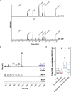
The β-1,3;1,6-glucan decasaccharide β-GD is released from the S. indica EPS matrix upon treatment with the barley apoplastic glycosyl hydrolase HvBGLUII. A, Glycosyl hydrolases specific for β-1,3;1,6-glucans were used for the characterization of the EPS matrix, CW, and β-GD. The β-1,3-endoglucanases from T. harzianum (TLE) and H. pomatia as well as FaGH17a and HvBGLUII are shown as open scissors (in blue). FaGH17a is represented as closed scissors because it does not hydrolyze glycosidic bonds of β-1,3-glucosyl residues substituted with β-1,6-glucosyl residues (in blue). FbGH30 is a β-1,6-exoglucanase (in orange). B, Analysis of digested EPS matrix or CW fractions by TLC. Several glucan fragments with different lengths are released from the EPS matrix and CW by the action of TLE and H. pomatia β-1,3-glucanase. HvBGLUII releases a glucan fraction from the β-glucan-containing EPS matrix but not from the CW. The experiment was repeated twice with Si EPS and Si CW isolated under different medium conditions (YPD and CM) and similar results were obtained. C, Analysis of digested EPS or CW fractions by MALDI-TOF mass spectrometry. The 1,661 Da β-GD corresponding to 10 hexoses is released from the EPS matrix but not from the CW of S. indica. The representative DP of hexoses is indicated on top of the m/z (M+Na)+ masses of oligosaccharides. The digestion of Si EPS with HvBGLUII was repeated independently more than three times with a similar result and the digestion of Si CW with HvBGLUII was performed two times with a similar result. D, 1H NMR spectrum of HPLC purified β-GD. E, Treatment of β-GD with various hydrolases followed by MALDI-TOF analysis of the products. The loss of three hexoses (−3 × 162 Da) as a result of treatment with FbGH30 is indicated with a dotted arrow. The experiment was performed two times with a similar result. F, Structure of the β-GD based on the 1H NMR spectrum. β-GD consists of a linear β-1,3-glucan backbone substituted with β-1,6-glucosyl moieties. Si, Serendipita indica; DP, degree of polymerization; Hexn, oligosaccharides with the indicated hexose composition; BC, backbone chain; TSC, terminal side-chain.
Figure 4
Figure 4
The β-GD released from S. indica EPS matrix scavenges ROS and enhances host colonization. A, Apoplastic ROS accumulation after treatment of barley roots of 8-day-old plants with 25-µM chitohexaose and/or purified β-GD from S. indica. ROS accumulation was monitored via a luminol‐based chemiluminescence assay. Treatment with Milli-Q water was used as mock control. Boxplot represents total ROS accumulation over the measured time period. Values represent mean ± sem from eight wells, each containing four root pieces. In total, roots from 16 individual barley plants were used per experiment. The assay was performed at least four times with independent β-GD preparations. Letters represent statistically significant differences in expression based on a one-way ANOVA and Tukey’s post hoc test (significance threshold: P ≤0.05). B, Prior to treatment of barley root pieces with the elicitors, β-GD was digested overnight (25°C, 500 rpm in heat block) with the glucanases FaGH17a and FbGH30, which led to complete digestion of β-GD (see also Figure 3E). As control, β-GD without the addition of enzymes (but instead with an equal volume of Milli-Q water) was treated similarly. Barley root pieces were treated with Milli-Q water (n = 16) and 25-µM Figure 4 (continued) chitohexaose alone (n = 16) or in combination with digested or undigested β-GD (300 µM, n = 12). The experiment was performed twice with similar results. Statistically significant differences are indicated by different letters based on a one-way ANOVA and Tukey’s post hoc test (significance threshold: P ≤0.05). C, Barley root pieces were collected 1 h after elicitor treatment and further processed for RNA extraction and cDNA synthesis. Expression changes of the elicitor-responsive gene HvWRKY2 were analyzed by RT-qPCR. Fold change expression were calculated by normalization to housekeeping gene expression (HvUBI) and mock treatment. Data from three independent experiments are indicated by different dot shapes. Letters represent statistically significant differences in expression based on two-way ANOVA (additive model, treatment + experiment) and Tukey’s post hoc test (significance threshold: P ≤0.05). Significant differences were associated with different treatments (F = 11.629, P = 1.58 × 10−6), but not with independent experiments (F = 2.227, P = 0.124). D, The capability of different carbohydrates to prevent hydrogen peroxide-based and horseradish peroxidase-catalyzed oxidation and precipitation of DAB was monitored. Respective sugars (or Milli-Q water as mock control) were pre-incubated with 1-mM H2O2 and 0.05-µM horseradish peroxidase before DAB (50 µM) was added. Scans of wells from 96-well plates were performed 16 h after DAB addition. The experiment was performed three times with similar results. E, Oxidative degradation of S. indica EPS matrix-derived β-GD (300 µM) by H2O2 was detected with an overnight Fenton reaction (1-mM H2O2, 100-µM FeSO4) followed by MALDI-TOF mass spectroscopic analysis. As controls, either sugar alone or the samples supplemented with 100-µM EDTA were used. F, Colonization of barley roots by S. indica upon daily application of sterile Milli-Q water (mock) or β-GD (100 or 300 µM). Fungal colonization in each biological replicate was assessed by RT-qPCR comparing the expression of the fungal housekeeping gene SiTEF and the plant gene HvUBI (n = 5–11). Boxplot elements in this figure: center line, median; box limits, upper and lower quartiles; whiskers, 1.5× interquartile range. Statistical significance was determined on the nontransformed values (before normalization to S. indica control treatment) using a two-tailed Student’s t test (*P ≤0.05). Si, Serendipita indica; RLU, relative light units; XXXG, xyloglucan heptasaccharide.
Figure 5
Figure 5
The β-GD derived from the hydrolysis of B. sorokiniana EPS matrix exhibits antioxidative properties. A, Glycosidic linkage analysis of B. sorokiniana EPS matrix and CW preparations. 2,3-hexopyranose, 2,3,4- hexopyranose, and 2,3,6- hexopyranose are abundant in the EPS matrix, whereas 4-glucose is abundant in the CW of B. sorokiniana. The experiment was performed with three independent biological replicates of Bs EPS and Bs CW and the TIC from one of the replicates is represented. B, Analysis of digested EPS or CW fractions by MALDI-TOF mass spectrometry. The 1,661-Da β-GD corresponding to 10 hexoses is released from the EPS matrix but not from the CW of B. sorokiniana. The representative DP of hexoses is indicated on top of the m/z (M+Na)+ masses of oligosaccharides. The digestion of Bs EPS with HvBGLUII was repeated independently three times with a similar result and the digestion of Bs CW with HvBGLUII was performed once. C, ROS burst assay was performed on barley roots treated with Milli-Q water (mock), chitohexaose (25 µM), Bs β-GD (300 µM), or a combination of chitohexaose and Bs β-GD. Boxplots represent total cumulative ROS accumulation over a measured time interval of 25 min. Each data point in the boxplot represents the integrated value from an individual well (center line, median; box limits, upper and lower quartiles; whiskers, 1.5× interquartile range). The experiment was performed three times with similar results. Statistically significant differences are indicated by different letters based on a one-way ANOVA and Tukey’s post hoc test (significance threshold: P ≤0.05). Bs, Bipolaris sorokiniana; DP, degree of polymerization; p, pyranose. aexact sugar moiety unknown; overrepresentation of linkages due to undermethylation cannot be excluded.
Figure 6
Figure 6
Model for the production and function of the conserved fungal EPS-derived β-1,3;1,6-glucan decasaccharide. The fungal-responsive GH17 family member HvBGLUII is found in the apoplast of barley roots and acts on β-1,3-glucan. Digestion of linear β-1,3-glucan (laminariheptaose) with HvBGLUII enhances ROS accumulation in barley roots, corroborating its role as a host defense enzyme with a function in β-glucan perception. To counteract the activity of HvBGLUII, plant-colonizing fungi produce a β-1,3;1,6-glucan-rich EPS matrix. The activity of HvBGLUII on the EPS matrix releases a conserved β-GD, which is resilient to further digestion by GH17 family members. The β-GD acts as a carbohydrate-class effector by scavenging ROS and enhancing fungal colonization. Lectins containing WSC domains are enriched in the outer EPS matrix and lectins containing LsyM domains are enriched in the CW of S. indica. Graphical illustration was designed with the BioRender online tool.

Comment in

References

    1. Adams EL, Rice PJ, Graves B, Ensley HE, Yu H, Brown GD, Gordon S, Monteiro MA, Papp-Szabo E, Lowman DW, et al. (2008) Differential high-affinity interaction of dectin-1 with natural or synthetic glucans is dependent upon primary structure and is influenced by polymer chain length and side-chain branching. J Pharmacol Exp Therap 325: 115–123 - PubMed
    1. Becker S, Scheffel A, Polz MF, Hehemann J-H (2017) Accurate quantification of laminarin in marine organic matter with enzymes from marine microbes. Appl Environ Microbiol 83: e03389–16 - PMC - PubMed
    1. Benaroudj N, Lee DH, Goldberg AL (2001) Trehalose accumulation during cellular stress protects cells and cellular proteins from damage by oxygen radicals. J Biol Chem 276: 24261–24267 - PubMed
    1. Boller T, Felix G (2009) A renaissance of elicitors: perception of microbe-associated molecular patterns and danger signals by pattern-recognition receptors. Annu Rev Plant Biol 60: 379–406 - PubMed
    1. Boulos S, Nystrom L (2017) Complementary sample preparation strategies for analysis of cereal beta-glucan oxidation products by UPLC-MS/MS. Front Chem 5: 90 - PMC - PubMed

Publication types