

Fluorogenic Cyclopropenones for Multicomponent, Real-Time Imaging
- PMID: 35442034
- PMCID: PMC9377832
- DOI: 10.1021/jacs.2c02058
Fluorogenic Cyclopropenones for Multicomponent, Real-Time Imaging
Abstract
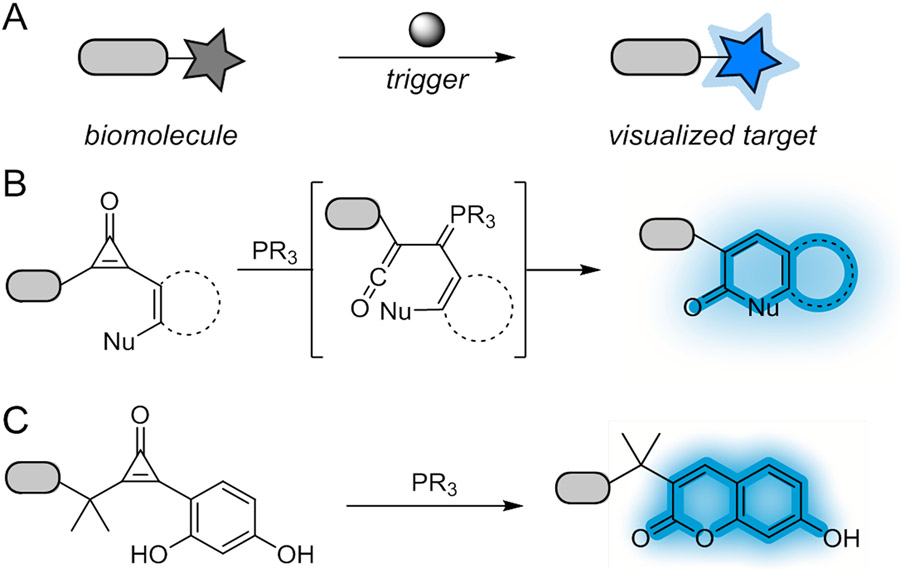
Fluorogenic bioorthogonal reactions enable biomolecule visualization in real time. These reactions comprise reporters that "light up" upon reaction with complementary partners. While the spectrum of fluorogenic chemistries is expanding, few transformations are compatible with live cells due to cross-reactivities or insufficient signal turn-on. To address the need for more suitable chemistries for cellular imaging, we developed a fluorogenic reaction featuring cyclopropenone reporters and phosphines. The transformation involves regioselective activation and cyclization of cyclopropenones to form coumarin products. With optimal probes, the reaction provides >1600-fold signal turn-on, one of the highest fluorescence enhancements reported to date. The bioorthogonal motifs were evaluated in vitro and in cells. The reaction was also found to be compatible with other common fluorogenic transformations, enabling multicomponent, real-time imaging. Collectively, these data suggest that the cyclopropenone-phosphine reaction will bolster efforts to track biomolecule targets in their native settings.
Figures

Similar articles
-
Constructing New Bioorthogonal Reagents and Reactions.Acc Chem Res. 2018 May 15;51(5):1073-1081. doi: 10.1021/acs.accounts.7b00606. Epub 2018 May 4. Acc Chem Res. 2018. PMID: 29727171 Free PMC article.
-
A Bioorthogonal Ligation of Cyclopropenones Mediated by Triarylphosphines.J Am Chem Soc. 2015 Aug 19;137(32):10036-9. doi: 10.1021/jacs.5b06969. Epub 2015 Aug 7. J Am Chem Soc. 2015. PMID: 26252114
-
Fluorogenic Coumarins Activated via Bioorthogonal Reactions.J Org Chem. 2025 Jul 25;90(29):10545-10549. doi: 10.1021/acs.joc.5c01117. Epub 2025 Jul 10. J Org Chem. 2025. PMID: 40639935
-
Bioorthogonal Reactions of Triarylphosphines and Related Analogues.Chem Rev. 2021 Jun 23;121(12):6802-6849. doi: 10.1021/acs.chemrev.1c00014. Epub 2021 Jun 8. Chem Rev. 2021. PMID: 34101453 Free PMC article. Review.
-
Molecular Design of Bioorthogonal Probes and Imaging Reagents Derived from Photofunctional Transition Metal Complexes.Acc Chem Res. 2020 Jan 21;53(1):32-44. doi: 10.1021/acs.accounts.9b00416. Epub 2020 Jan 9. Acc Chem Res. 2020. PMID: 31916746 Review.
Cited by
-
Platform for Orthogonal N-Cysteine-Specific Protein Modification Enabled by Cyclopropenone Reagents.J Am Chem Soc. 2022 Jun 15;144(23):10396-10406. doi: 10.1021/jacs.2c02185. Epub 2022 Jun 5. J Am Chem Soc. 2022. PMID: 35658467 Free PMC article.
-
Bioorthogonal cyclopropenones for investigating RNA structure.bioRxiv [Preprint]. 2024 Oct 24:2024.10.22.619649. doi: 10.1101/2024.10.22.619649. bioRxiv. 2024. Update in: ACS Chem Biol. 2024 Dec 20;19(12):2406-2411. doi: 10.1021/acschembio.4c00633. PMID: 39484557 Free PMC article. Updated. Preprint.
-
Bioorthogonal Cyclopropenones for Investigating RNA Structure.ACS Chem Biol. 2024 Dec 20;19(12):2406-2411. doi: 10.1021/acschembio.4c00633. Epub 2024 Dec 6. ACS Chem Biol. 2024. PMID: 39641920 Free PMC article.
-
Recent Advances in Bioorthogonal Ligation and Bioconjugation.Top Curr Chem (Cham). 2023 Nov 22;381(6):35. doi: 10.1007/s41061-023-00445-6. Top Curr Chem (Cham). 2023. PMID: 37991570 Free PMC article. Review.
-
Supramolecular Guest Exchange in Cucurbit[7]uril for Bioorthogonal Fluorogenic Imaging across the Visible Spectrum.ACS Cent Sci. 2024 Oct 8;10(10):1945-1959. doi: 10.1021/acscentsci.4c01080. eCollection 2024 Oct 23. ACS Cent Sci. 2024. PMID: 39463826 Free PMC article.
References
-
- van de Linde S; Heilemann M; Sauer M Live-Cell Super-Resolution Imaging with Synthetic Fluorophores. Annu. Rev. Phys. Chem 2012, 63, 519–540. - PubMed
-
- Specht EA; Braselmann E; Palmer AE A Critical and Comparative Review of Fluorescent Tools for Live-Cell Imaging. Annu. Rev. Physiol 2017, 79, 93–117. - PubMed
